
Special Article - Surgical Case Reports
Austin J Surg. 2015;2(3): 1057.
GAsymptomatic Ulnar Nerve Neurilemmoma Masquerading as a Lipoma
Wani Bhushan N¹* and Jajoo Suhas N²
¹Department of Surgery, MVPS DVP Medical College,India
²Department of Radiodiagnosis, Jawaharlal Nehru Medical College, India
*Corresponding author: Wani Bhushan N,Department of Surgery, MVPS’s Dr. Vasantrao Pawar Medical College, Nashik, India
Received: November 24, 2014; Accepted: March 18, 2015;Published: April 02, 2015
Abstract
Peripheral nerve tumours are rare, the commonest being the (benign) neurinoma which usually presents with mild symptoms or even no neurological deficit. Here in our case, patient came with mild, diffuse mass and discomfort. The clinical neurological examination was not conclusive. Asymptomatic unexpected neurilemmoma of the ulnar nerve was excised considering it as lipoma. After surgery its nervous origin was confirmed and the histopathological examination confirmed the diagnosis of neurilemmoma. Point to remember is that, even a surface lesion that appears benign, it should be seriously considered peripheral nerve sheath tumours as a possible differential diagnosis.
Keywords: Neurilemmoma; Schwannoma; Neurofibroma; Lipoma
Introduction
Peripheral nerve tumours are rare, the commonest being the (benign) neurinoma [1]. In 1910 Verocay had first time postulated that neurinoma has histologically to be distinguished from neurofibromas [2] and it also arises from Schwann cells [3]. In the literature the tumor is called Neurilemmoma or Schwannoma (because of its origin). They can present with mild symptoms or even no neurologic deficit, so difficulty in diagnosis clinically is common and they have been confused with lipomas, haemangiomas, neurofibromas, etc. [1,3]. Careful dissection is of paramount important during the surgery, to prevent unfortunate resection of the nerve. An ample degree of attention is necessary for the inclusion of peripheral nerve tumours as a differential diagnosis of an upper extremity mass [3].
Case
A 38 years old man presented with a swelling over posterior compartment of the arm. It was first noticed 4-5 years ago after he had had an injury over the area, but lately it has been associated with dull pain and discomfort (Figure 1). He had no significant past medical or family history. There were no other systemic symptoms. On examination, there was a non-tender, firm, deep swelling, free from the skin with restricted mobility in vertical axis over the extensor compartment of the arm. The surrounding skin was normal without any scars or pigmented areas. The clinical neurological examination was without any deficit and not conclusive. No biopsy was undertaken before definitive surgery, considering benignity at clinical and imaging examination of swelling.
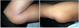
Figure 1: Clinical Photograph of swelling over posterior compartment of arm.
The patient underwent surgical excision of the mass performed with a posteriomedial approach. The mass was located eccentrically within the epineurium of the ulnar nerve. It was carefully dissected from the nerve without any untoward incident (Figure 2A). It was a well-encapsulated, yellowish-white, firm nodule, without any pedicle from or adjacent vessels (Figure 2B). The mass was removed in to, haemostasis achieved and a primary closure was performed. Gross pathologic evaluation demonstrated a well-circumscribed, xanthochromic, rubbery mass with an appearance that suggested of lipoma. Microscopic evaluation confirmed the diagnosis of schwannoma, demonstrating densely cellular area, arranged in short bundles of interlacing fasicles and adjacent nerve fibres (Figure 3).
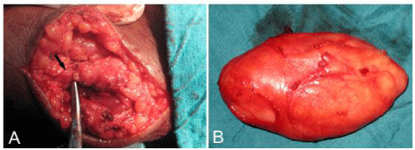
    Figure 2:  Operative photograph [A] Carefully dissected mass located within
the epineurium of theulnar nerve [B] Well-encapsulated, yellowish-white, firm
nodule, without any pedicle from or adjacent vessels.
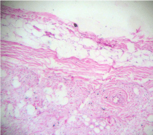
    Figure 3:  Photomicrograph reveals that the tumor consists of focal cellular
‘Antoni A’ areas and hypocellular ‘Antoni B’ areas with vacuolar degeneration.
Discussion
Neurilemmoma (schwannoma) arising from the Schwann cells of the nervous system constitutes about 5% of all benign soft-tissue tumours [1]. They are slightly preponderant in men than women, with peak occurrence between 30 and 60 years of age [2]. They are usually solitary, circumscribed, and encapsulated tumours eccentrically located on proximal nerves or spinal nerve roots. They are usually found on the flexor surface of the forearm and hand, and multiple tumours can be located within a single major nerve, its branches, or both [4].
Their usual presentation as, painless mass with paresthesi as and positive Tinel’s sign over the nerve with exceptions if the nerve is in a restricted space such as carpal or tarsal tunnel. Diagnostic suspect of schwannoma is made preoperatively on imaging studies like ultrasounds, MRI which could be helpful in differential diagnosis. The suspect may be confirmed by frozen section; but in our case, macroscopic aspect of the lesion was that of a benign lipoma; so frozen sections was not done before excision. Usually final diagnosis is made intraoperatively or postoperatively and confirmed on histopathology with the chance to compromise the outcome of the treatment. Sometimes in large nerve involvement, the mass is characteristically eccentric with respect to the affected nerve, and the nerve displaced to the periphery of the mass; in contrast to small nerves. A schwannoma has a true capsule composed of epineurium; [5] thus successful resections are possible, as growth of schwannoma within the epineurium creates encapsulation. In contrast, neurofibroma arising from the nerve fascicle is centrally located, rarely encapsulated, and cannot be separated from the involved nerve [6].
The malignant peripheral nerve sheath tumours, account for approximately 6% of all sarcomas, with approximately one-half occurring in the setting of neurofibromatosis [1,5]. Benign soft-tissue tumours are 100 times more frequent than sarcomas; thus, malignant peripheral nerve sheath tumours are relatively uncommon statistically. As like to schwannoma, malignant peripheral nerve sheath tumours usually arise from major nerve trunks (in the proximal extremities and torso), often containing prominent areas of haemorrhage and necrosis [2,6,7]. Although they have no specific imaging features and high-gradation, their aggressive biologic behavior may be suggested by indistinct margins, the infiltrative nature of the lesion within the nerve and adjacent structures, and lesion heterogeneity [7]; which requires an excision with wide or radical surgical margins and the resection of the nerve trunk of origin.
Conclusion
When a clinician encounters a surface lesion that appears benign, he or she should seriously consider peripheral nerve sheath tumours as a possible differential diagnosis.
References
- Morris JH. The nervous system. In: Cotran RS, Kumar V, Robbins SL, editors. Robbins Pathologic Basis of Disease. 4th edn. Philadelphia, WB Saunders. 1989; 1445-1447.
- Sandberg K, Nilsson J, Soe Nielsen N, Dahlin LB. Tumours of peripheral nerves in the upper extremity: a 22-year epidemiological study. Scand J Plast Reconstr Surg Hand Surg. 2009; 43: 43-49.
- Anthony DC, Vogel S. Peripheral nervous system. In: Damjanov I, Linder J, Anderson WAD, editors. Anderson's Pathology, 10th edn. St Louis, Mosby. 1996; 2824-2826.
- Vigler M, Levine LJ, Posner MA. Multiple neurilemomas in the upper extremity: a series of three cases. Bull NYU Hosp Jt Dis. 2008; 66: 61-64.
- Takase K, Yamamoto K, Imakiire A. Clinical pathology and therapeutic results of neurilemmoma in the upper extremity. J Orthop Surg. 2004; 12: 222-225.
- Classen DA, Rees H. Neurilemoma of the ulnar nerve. Can J Surg. 1996; 39: 356-357.
- Stout AP. The peripheral manifestations of the specific nerve sheath tumor (neurilemmoma). Am J Cancer. 1935; 24: 751-796.