
Special Article - Surgical Case Reports
Austin J Surg. 2015;2(6): 1071.
Patellar Clunk Syndrome after Patellofemoral Arthroplasty: A Case Report
Jason Wong*, Sunjin Kim and Jonathan B Courtney
Montefiore Medical Center, USA
*Corresponding author: Jason Wong, Montefiore Medical Center, USA
Received: May 29, 2015; Accepted: August 17, 2015; Published: August 25, 2015
Abstract
Patellar clunk syndrome is defined as a painful catching of the patella upon knee extension from a flexed position after total knee arthroplasty. It is usually caused by the formation of a discrete fibro synovial nodule at the junction between the superior pole of patella and the quadriceps tendon. We report a case of patellar clunk syndrome in a 51 year-old female after a patellofemoral joint replacement that improved after arthroscopic nodule excision and lateral retinacular release.
Case Report
A 51 year-old female presented to the osteoarthritis clinic with a chief complaint of anterior left knee pain. The pain was exacerbated with stairs, squatting, and getting up from a seated position. On exam, she had normal patella tracking, full knee range of motion, and positive patella grind. Radiographs of the left knee revealed large lateral osteophyte on the patella with joint space narrowing consistent with patella-femoral osteoarthritis (Figures 1A-1C).

Figure 1a: AP of the left knee.

Figure 1b: Lateral of the left knee.

Figure 1c: Axial view of the left knee.
She underwent over a year of non-operative management, including nonsteroidal anti-inflammatory drugs (NSAIDs), physical therapy, as well as intra-articular steroid injections. Over time, these modalities failed to provide her meaningful relief. She had undergone a right patella-femoral arthroplasty two years previously for isolated patella-femoral arthritis, and had an excellent post-operative outcome. Given that she had failed conservative management of her left patella-femoral arthritis, she was indicated for a left Patella- Femoral Arthroplasty (PFA).
The surgery was performed with a tourniquet set to 300mmHg. An anterior longitudinal incision was made centered over the patella, followed by a standard medial parapatellar arthrotomy. The medial and lateral compartments were inspected, and found to have relatively normal cartilage, and the decision was made to proceed with the PFA. An intramedullary guide was used, and the femoral external rotation was set parallel to the intercondylar line as well as perpendicular to Whiteside’s line, a perpendicular line to the axis of the center of the trochlea and the intercondylar notch [1]. Using the system’s cutting guides, the appropriate femoral cuts were made. After measuring the thickness of the patella, the appropriate patella cut was made to subchondral bone. The prosthetic trials were placed, and it was noted that the femoral trial was flush with the surrounding articular cartilage. In taking the knee through a full range of motion, there was excellent patella tracking throughout, without catching or lateral subluxation. Therefore, the decision was made not to perform a lateral release. The trials were removed, the wound was irrigated and dried, and the actual components (Stryker Avon #29 Patella button with an Avon extra small patella-femoral component) were cemented into place. When the cement had fully cured, the knee was taken through a range of motion, and again, it was noted to have excellent patella tracking without catching or lateral subluxation. The athrotomy was closed with interrupted sutures, without a medial advancement. The knee was sequentially ranged to ensure proper tracking. The remaining closure of the subcutaneous layer and skin were performed as per normal protocol for a total knee replacement. The tourniquet was deflated after a sterile, compressive dressing was applied.
Post-operatively, the patient participated in physical therapy, working on her range of motion, quadriceps strengthening, and gait training. At the six-week visit, her pain was much improved. Her active range-of-motion of her knee was 0-120° with normal patella tracking. X-rays taken showed her components were in appropriate position (Figures 2A-2C). She returned for follow-up roughly six months after surgery, complaining of a snapping sensation in her knee. She stated that for the past two months, after walking one or two blocks, the snapping would occur when she bent her knee during normal gait. On exam, the incision was well-healed. There was a palpable snapping sensation over the superolateral aspect of the patella that occurred when the patient actively moved her knees at 45° from flexion to extension. Patella tracking was otherwise normal. This was very bothersome for her, and she opted for arthroscopic management for presumed diagnosis of “patella clunk”.

Figure 2a: AP of the left knee status post PFA.
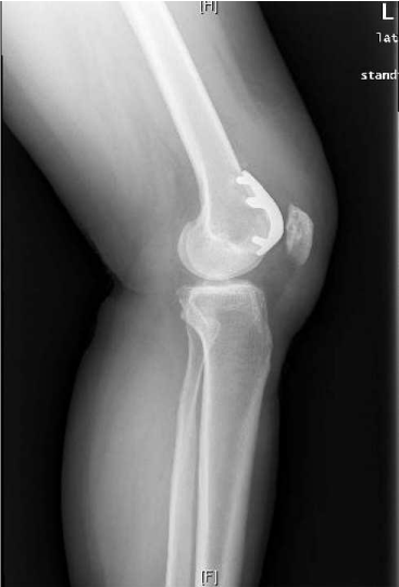
Figure 2b: Lateral of the left knee status post PFA.
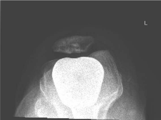
Figure 2c: Axial view of the left knee status post PFA.
Shortly thereafter, an arthroscopy was performed. Immediately noted was a fibrous nodule located at the supero-lateral margin of the patellar button (Figure 3). The remaining cartilage and intra-articular structures were intact. The nodule was excised with electrosurgical ablation (Figures 4A and 4B). A lateral directed force was applied to the patella, and the lateral retinacular structures were deemed to be tight. Therefore, a lateral release was performed as well.
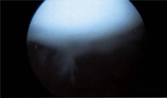
Figure 3: Fibrous nodule at the superior aspect of the patella.
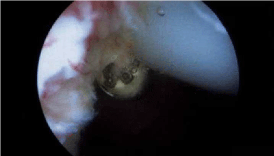
Figure 4a: Fibrous nodule being cauterized.
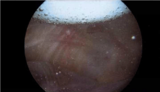
Figure 4b: Fibrous nodule is removed.
Post-operatively, the patient did quite well. Her snapping sensation was gone. Her knee pain was gone. She had no complaints from her knee up to the last clinic visit.
Discussion
Patella clunk syndrome is a relatively common cause for unsatisfactory results after Total Knee Arthroplasty (TKA) [2]. John Insall described a case of anterior knee pain associated with a peripatellar fibrous nodule that resolved following operative resection [3]. The term “Patella Clunk” was coined by Hozack [4] in a series of patients who had undergone posterior-stabilized TKA, and was defined as a painful catching, grinding, or jumping of the patella upon knee extension from a flexed position. It is usually caused by a discrete fibro synovial nodule being trapped within the intercondylar box of the Posterior Stabilizing (PS) femoral component during knee flexion. When the knee is extended to about 30 to 35 degrees, the nodule dislodges resulting in an audible and often painful clunk. As a result, patella clunk syndrome is most commonly associated with PS knees with an incidence of 0 to 18 percent; but it has also been associated with Cruciate Retaining (CR) total knee arthroplasty as well [3,5-11]. In the English-language literature, there has only been one case report on patella clunk syndrome that was associated with patella-femoral replacement [12]. The current report will however be the first report to include intraoperative images in the publication as well as providing explanations of patella clunk syndrome specific to PFA.
There are several theories to the etiology of patella-clunk syndrome in TKA. Femoral component geometry, thicker tibial inserts, anterior tibial tray placement, and flexed femoral components have all been implicated as possible [13]. In PFA, however, these considerations do not apply. In their report of patellar clunk syndrome in PFA, Sringari et al suggested that the transition point of metallic implant to the articular cartilage of the femoral component acted like the notch and femoral groove as in a posterior stabilized TKA, thus catching and causing the clunk [12].
We performed an arthroscopic debridement of the nodule (Figures 5 & 6), followed by a lateral retinacular release. Upon releasing the retinaculum, the tissue spread roughly 1cm indicating that it was under tension. It has been shown that the contact force of the patella-femoral joint is significantly reduced followed lateral release [14]. Many patients with isolated patella-femoral arthritis do have lateral mal-tracking with tight retinacular structures. We suspect that our patient had a tight lateral retinaculum, causing increased pressure at the patella-femoral joint, which in turn, increased the stress at the metallic implant-articular cartilage junction, thus causing the fibrous nodule to form at the bone-implant interface.
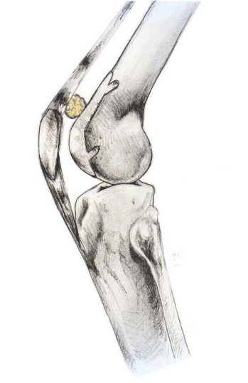
Figure 5: Illustration of fibrous nodule when knee in extension.
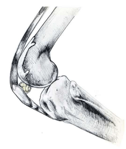
Figure 11: Illustration of fibrous nodule when knee in flexion.
There is no consensus regarding lateral release in PFA compared to the well-published indications of lateral release in total knee arthroplasty [15]. There have been several reports with an incidence of lateral release ranging from 27% to 88% [16-19]. There is no data regarding the effect of the lateral release on patella-femoral contact forces in PFA. However, based on the previously described research, one would intuitively expect that the forces would decrease. In this case, we believe that the increased force at the patella-femoral contact point after the PFA secondary to tight lateral retinaculum may have led to the formation of a fibrous nodule in our patient. She felt immediate improvement after the arthroscopic nodule excision and lateral release, and continues to enjoy a pain-free knee at her last follow up visit.
In conclusion, we have described a case of patella clunk syndrome following PFA, which may have been attributed to abnormal patellofemoral biomechanics that has resolved with arthroscopic excision with lateral retinacular release.
Acknowledgement
We would like to thank Ms. Rachel Ho for providing us with the illustrations.
References
- Poilvache PL, Insall JN, Scuderi GR, Font-Rodriguez DE. Rotational landmarks and sizing of the distal femur in total knee arthroplasty. Clin Orthop Relat Res. 1996; 35-46.
- Conrad DN, Dennis DA. Patellofemoral crepitus after total knee arthroplasty: etiology and preventive measures. Clin Orthop Surg. 2014; 6: 9-19.
- Insall JN, Lachiewicz PF, Burstein AH. The posterior stabilized condylar prosthesis: a modification of the total condylar design. Two to four-year clinical experience. J Bone Joint Surg Am. 1982; 64: 1317-1323.
- Hozack WJ, Rothman RH, Booth RE Jr, Balderston RA. The patellar clunk syndrome. A complication of posterior stabilized total knee arthroplasty. Clin Orthop Relat Res. 1989; 203-208.
- Meftah M, Ranawat AS, Ranawat CS. The natural history of anterior knee pain in 2 posterior-stabilized, modular total knee arthroplasty designs. J Arthroplasty. 2011; 26: 1145-1148.
- Beight JL, Yao B, Hozack WJ, Hearn SL, Booth RE Jr. The patellar "clunk" syndrome after posterior stabilized total knee arthroplasty. Clin Orthop Relat Res. 1994; 139-142.
- Clarke HD, Fuchs R, Scuderi GR, Mills EL, Scott WN, Insall JN. The influence of femoral component design in the elimination of patellar clunk in posterior-stabilized total knee arthroplasty. J Arthroplasty. 2006; 21: 167-171.
- Fukunaga K, Kobayashi A, Minoda Y, Iwaki H, Hashimoto Y, Takaoka K. The incidence of the patellar clunk syndrome in a recently designed mobile-bearing posteriorly stabilised total knee replacement. J Bone Joint Surg Br. 2009; 91: 463-468.
- Lonner JH, Jasko JG, Bezwada HP, Nazarian DG, Booth RE Jr. Incidence of patellar clunk with a modern posterior-stabilized knee design. Am J Orthop (Belle Mead NJ). 2007; 36: 550-553.
- Yau WP, Wong JW, Chiu KY, Ng TP, Tang WM. Patellar clunk syndrome after posterior stabilized total knee arthroplasty. J Arthroplasty. 2003; 18: 1023-1028.
- Pollock DC, Ammeen DJ, Engh GA. Synovial entrapment: a complication of posterior stabilized total knee arthroplasty. J Bone Joint Surg Am. 2002; 84-84A: 2174-2178.
- Sringari T, Maheswaran SS. Patellar clunk syndrome in patellofemoral arthroplasty- a case report. Knee. 2005; 12: 456-457.
- Costanzo JA, Aynardi MC, Peters JD, Kopolovich DM, Purtill JJ. Patellar clunk syndrome after total knee arthroplasty; risk factors and functional outcomes of arthroscopic treatment. J Arthroplasty. 2014; 29: 201-204.
- Clifton R, Ng CY, Nutton RW. What is the role of lateral retinacular release? J Bone Joint Surg Br. 2010; 92: 1-6.
- Diduch DR, Scuderi GR, Scott WN, Insall JN, Kelly MA. The efficacy of arthroscopy following total knee replacement. Arthroscopy. 1997; 13: 166-171.
- Arciero RA, Toomey HE. Patellofemoral arthroplasty. A three- to nine-year follow-up study. Clin Orthop Relat Res. 1988; 60-71.
- Cartier P, Sanouiller JL, Grelsamer R. Patellofemoral arthroplasty. 2-12-year follow-up study. J Arthroplasty. 1990; 5: 49-55.
- Tauro B, Ackroyd CE, Newman JH, Shah NA. The Lubinus patellofemoral arthroplasty. A five- to ten-year prospective study. J Bone Joint Surg Br. 2001; 83: 696-701.
- Krajca-Radcliffe JB, Coker TP. Patellofemoral arthroplasty. A 2- to 18-year followup study. Clin Orthop Relat Res. 1996; 143-151.