Special Article - Surgical Case Reports
Austin J Surg. 2015; 2(6): 1072.
Strangulated Internal Hernia through the Lesser Sac – An Unusual Cause of Small Bowel Obstruction
Sangram Keshari Panda¹* and Amita Panda²
¹Department of General Surgery, Kalinga Institute of Medical Science, India
²M.K.C.G Medical College, India
*Corresponding author: Sangram Keshari Panda, Senior Resident, Department of General Surgery, Bhubaneswar, Odisha, India
Received: August 17, 2015; Accepted: September 21, 2015; Published: September 25, 2015
Abstract
Introduction: The incidence of internal hernias is 0.2% to 2%. It is a rare cause of intestinal obstruction and leads from 0.5 to 4.1% of acute obstruction cases caused by hernia.
Presentation of Case: We report a case of internal hernia traversing the lesser sac with a primary defect in the greater omentum and a second defect in the lesser omentum, causing small bowel obstruction which is extremely rare.
Discussion: The most common causes of small bowel obstruction in adults are adhesions bands, malignancy and hernias. Internal Hernia (IH) is defined as herniation of viscera through a normal or abnormal aperture within the peritoneal cavity. Internal hernias are infrequent, accounting for 0.2 to 0.9% of the cases of intestinal obstruction. Transomental hernias through the greater or lesser omentum are even rarer, representing 1 to 4% of all internal hernias, with hernias occurring through both the omentum being extremely rare as in our case.
Conclusion: When a case of acute intestinal obstruction is reported, after ruling out the known causes of obstruction, surgeon should have high suspicion of internal hernia to reduce the risk of intestinal ischemia, necrosis and perforation; thus decreasing the postoperative morbidity and mortality.
Keywords: Strangulated internal hernia; Intestinal obstruction; Absolute constipation; Lesser sac
Introduction
An internal hernia is defined as the protrusion of a viscous through a normal or abnormal opening within the confines of the abdominal cavity [1]. The classification is based on anatomic regions where internal hernias with distinctive clinical and radiographic features occur: paraduodenal (left > right) (53%), foramen of Winslow (8%), pericecal (13%), Inter sigmoid (6%), Transmesenteric (8%), Transomental (1–4%), Retroanastomotic, Supravesical and pelvic (6%) [2]. The incidence of internal hernias is 0.2% to 2%. It is a rare cause of intestinal obstruction and leads from 0.5 to 4.1% of acute obstruction cases caused by hernia [3]. We report a case of internal hernia traversing the lesser sac with a primary defect in the greater omentum and a second defect in the lesser omentum, causing small bowel obstruction which is extremely rare.
Case Report
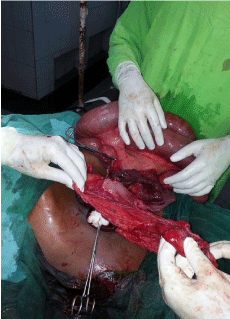
A 32 year old male patient was admitted to emergency department of SCB Medical College & hospital, Cuttack with complaints of repeated episodes of vomiting, absolute constipation, colicky abdominal pain and abdominal distension of duration two days. He had no past history of tuberculosis or any abdominal surgery. There is no similar incidence in the past. Upon clinical examination, patient was dehydrated and afebrile with a pulse rate of 96/min and a blood pressure of 102/ 58 mm of Hg. On abdominal examination, abdomen was distended, non tender, no visible peristalsis and bowel sounds were absent. On digital rectal examination, rectum was normal. Hematological and biochemical investigations were within normal limit. Plain X-ray abdomen showed multiple air fluid levels. Abdominal ultra sonography was performed which did not reveal any significant information. A nasogastric tube was inserted. With a diagnosis of acute intestinal obstruction, the patient underwent an exploratory laparotomy that revealed about 90-cm proximal ileal segment herniated through an opening in the greater omentum (Figure 1) and came out through the second opening in the lesser omentum after traversing the lesser sac. The ileal segment was strangulated in the secondary defect in the lesser omentum (Figure 2). Upon widening the hernial orifice and resecting both ends of the strangulated bowel, the ischemic bowel was removed and a primary anastomosis was performed. The hernial defects were repaired. The postoperative course was uneventful and the patient was discharged on 10th postoperative day. He was followed up as an outpatient for 3 months and had no further difficulties.

    Figure 1:  Herniation of proximal part of small intestine through a defect in
greater omentum.

    Figure 2:  Herniated gangrenous small bowel through the lesser omentum
after traversing the lesser sac.
Discussion
The most common causes of Small bowel obstruction in adults are adhesions bands, malignancy and hernias [4]. Congenital hernias occur most commonly in infancy and childhood but rarely can be seen in adults. Internal Hernia (IH) is defined as herniation of viscera through a normal or abnormal aperture within the peritoneal cavity. It has been demonstrated that internal hernia often remain undiagnosed before emergency laparotomy since the symptoms of IH may be nonspecific ranging from mild abdominal discomfort to sudden onset intestinal obstruction. Furthermore, it occasionally leads to gangrene necessitating bowel resection of varying extent which may contribute to high mortality.
Transomental hernias through the greater or lesser omentum are even rarer, representing 1 to 4% of all internal hernias, with hernias occurring through the lesser omentum being extremely rare as in our case. Predisposing factors for Transomental hernias include congenital anatomic defects of the liver, lesser sac, mesentery, as well as the presence of adhesions or increased intra-abdominal pressure. Abnormal Transomental openings are usually congenital, and rarely traumatic or iatrogenic [5].
Petersen’s space Hernia is an internal hernia, arising after any type of gastrojejunostomy (most frequently after Roux-en-Y anastomosis) and its boundaries are now a days described as the transverse mesocolon, the retro peritoneum and the Roux limb mesentery. One major challenge with these patients is that the presenting signs, symptoms and radiological examinations may be nonspecific or non diagnostic. Though Petersen’s space hernia is a forgotten diagnosis for most of surgeons in the last 30 years due to the diminished frequency of gastrojejunostomy, the exponential growth of laparoscopic gastric bypass for the treatment of morbid obesity increasingly bring this kind of complication [6].
The literature on internal hernia [7-9] published during last decade emphasizes that a hypothesis of internal hernia should be considered for patient with signs and symptoms of intestinal obstruction, particularly in absence of inflammatory intestinal disease, external hernia or previous laparotomy. There is extreme difficulty in making diagnosis of internal hernia preoperatively whether incarcerated or strangulated. The overall management of acute intestinal obstruction requires appropriate initial resuscitation and naso-gastric decompression, followed by immediately laparotomy. A median laparotomy incision, followed by upper or lower extension when needed is usually adequate for accessing the unpredictable site of obstruction and for any procedure required. A finding of herniation through an intraperitoneal orifice makes a diagnosis of internal hernia. Next a careful examination of abdominal cavity is done to identify other structures involved. The surgical management of internal hernia includes reduction of herniated structures, resection of ischaemic intestinal segments and closure of hernial orifice. Widening of the orifice has the risk of vascular lesions which can be avoided through knowledge of anatomy of peritoneal fossa and the regional vessels. Mild ischaemia is reverted back a few minutes after the loop is released. In the presence of necrosis, perforation or irreversible ischaemia intestinal resection is performed. Closure of hernial orifice is generally indicated for prevention of recurrence of hernia through abnormal orifice [9].
Conclusion
When a case of acute intestinal obstruction is reported after ruling out the known causes of obstruction a hypothesis of internal hernia can be made. The task on exploration is to confirm the provisional diagnosis of internal hernia. The policy of a surgeon is to have a suspicion of internal hernia reducing the risk of intestinal ischemia, necrosis and perforation thus decreasing the postoperative morbidity and mortality. So rapid and proper evaluation and immediate therapy is mandated in cases of small bowel obstruction with suspicion of internal hernia.
References
- Uchiyama S, Imamura N, Hidaka H, Maehara N, Nagaike K, Ikenaga N, et al. An unusual variant of a left par duodenal hernia diagnosed and treated by laparoscopic surgery: report of a case. Surg Today. 2009; 39: 533-535.
- Gomes R, Rodrigues J. Spontaneous adult transmesenteric hernia with bowel gangrene. Hernia. 2011; 15: 343-345.
- Meyers MA. Para duodenal hernias: radiologic and arteriographic diagnosis. Radiology. 1970; 95: 29-37.
- Foster NM, McGory ML, Zingmond DS, Ko CY. Small bowel obstruction: a population-based appraisal. J Am Coll Surg. 2006; 203: 170-176.
- Gulino D, Giordano O, Gulino E. Les hernies interns de l 'abdomen. À propos de 14 cas. J Chir (Paris) 1993; 130: 179-195.
- Faria G, Preto J, Oliveira M, Pimenta T, Baptista M, Costa-Maia J. Petersen's space hernia: A rare but expanding diagnosis. Int J Surg Case Rep. 2011; 2: 141-143.
- Pessaux P, Tuech JJ, Derouet N, Plessis R, Roncerray J, Arnaud JP. Internal hernia: a rare cause of intestinal obstruction. Apropos of 14 cases. Nan Chir. 1999: 53; 870-873.
- Takagy Y, Yasuda K, Nakada T, Abe T, Matsuura H, Saji S. A case of strangulated transomental hernia diagnosed preoperatively. Am J Gastroent. 1996; 91: 1659-1661.
- Ozenc A, Ozdemir A, Coskun T. Internal hernia in adults. Int Surg. 1998; 83: 167-170.