Special Article - Surgical Case Reports
Austin J Surg. 2015; 2(7): 1076.
Pulmonary Sequestration with Rare Blood Supply in an Asymptomatic Adult Female
Nieh CC1,2*, Chua YC¹ and Agasthian T¹
¹Department of Cardiothoracic and Vascular Surgery, National University Hospital, Singapore
²Yong Loo Lin School of Medicine, National University of Singapore, Singapore
*Corresponding author: Nieh CC, Department of Cardiothoracic and Vascular Surgery, National University Hospital, Singapore
Received: September 28, 2015; Accepted: November 19, 2015; Published: November 26, 2015
Abstract
Intralobar pulmonary sequestration with two aberrant vessels connected to aorta is very rare. We describe the case of a 61-year-oldasymptomatic Chinese female with an incidental finding of abnormal chest radiograph showing a left lower lobe mass. Computed Tomography scan of the Thorax (CT Thorax) revealed a cystic mass in the left lower lobe, supplied by two branches from the thoracic aorta, likely to be intra lobar pulmonary sequestration. A dystrophic bronchial tree that did not communicate with the normal bronchial tree was identified. The sequestered lung was completely excised via Video-Assisted Thoracoscopic Surgery (VATS).
The patient had an uncomplicated clinical course after surgery and was discharged on postoperative-day three in satisfactory condition. Histology reviewed mild chronic inflammation along with overlying interstitial fibrosis and bronchial metaplasia. In addition, bronchial plugging along with an abundant arterial supply was identified to be consistent with intralobar pulmonary sequestration
CT Thorax is ideal to visualise the vascular anatomy which is essential for surgical management. Were commend that surgical segmental resection with VATS should continue to be the standard of care in asymptomatic adult patients with this rare condition.
Keywords: Pulmonary sequestration; Thoracoscopic; Case report; Asymptomatic; Adult
Introduction
Pulmonary sequestration is a rare condition contributing to 0.15-6.4%ofallcongenitalmalformations of the lower respiratory tract [1,2]. Typically, it comprises of systemic arterial supply, usually derived from an aberrant branch of the lower thoracic or upper abdominal aorta, to an associated anomalous lung segment with variations in its venous drainage, usually to the left atrium, sometimes right atrium, vena cava, or azygous systems. These tissues are not connected to the trachea bronchial tree. Sequestrations are typically classified an atomically into intralobar and extra lobar forms, with the former being defined as a lung segment contained within the native pleural lining while the latter is contained in its own pleural investment.
Amongst both forms of sequestrations, intralobar type is the commonest, approximately 70 to 95 percent of all sequestrations. Sixty percent of intrapulmonary sequestrations are diagnosed within the first decade of life and are more common in male’s by a 3:1 ratio1. Symptoms are variable, depending on the anatomical location of the lesion, but usually related to chronic respiratory infection although sequestrations can be incidental findings on radiological investigations.
The exact pathogenesis of pulmonary sequestration is unclear, for which several theories have been suggested to elucidate the embryologic mechanism. There is another hypothesis that describes a disruption during the processes of lung development which distorts its arterial blood supply which subsequently leads to retention and proliferation of the nascent systemic capillary network [3,4]. Till now, no one is certain about the embryological development of this condition. Usual age of presentation for intra pulmonary sequestration and extra pulmonary sequestration are adolescence with signs and symptoms of recurrent infection, and infancy with respiratory failure respectively.
We now demonstrate a recent case of intralobar pulmonary sequestration supplied by two aberrant vessels arising from the aorta at our institution who presented at an age of 61withoutsymptoms.
Case
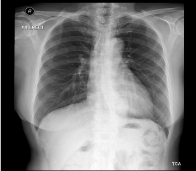
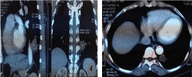
The patient is a sixty-one year old Chinese female with no significant past medical history. Computed tomography of Thorax (Figures 1 & 2) revealed a cystic mass in the left lower lobe, supplied by two solitary branches from the thoracic aorta likely intra lobar pulmonary sequestration. There was no pleural effusion or consolidation. A dystrophic bronchial tree that did not communicate with the normal bronchial tree was identified. Chest radiography showed a left lower lobe mass.

    Figure 1:  Chest radiography in the patient showing the lesion in left lower
lobe.

Figure 2: CT thoraxpre-operatively.
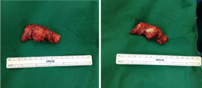
Upon admission, vitals were normal. On examination, she was well. The patient underwent elective VATS. The aberrant arterial supply from the thoracic aorta was identified after mobilization of the intra pulmonary sequestration (Figure 3). The artery was then ligated with the use of silk suture and surgical clips. The remainder of the segmentectomy was performed in the usual fashion via stapler.

Figure 3: Specimen of intra pulmonary sequestration.
A chest tube was inserted post-operatively and removed on 1st post-operative day.
The patient was discharged on post operative day three. Histology reviewed mild chronic inflammation along with overlying interstitial fibrosis and bronchial metaplasia. Bronchial pluggings along with two aberrant arterial supplies were identified to be consistent with intra lobar pulmonary sequestration. The patient has continued to progress well.
Discussion
Intra lobar pulmonary sequestration is a rare congenital anomaly with few reports of cases being diagnosed during adulthood. Patients can present with an incidental lesion in the lung on imaging asymptomatically. They may manifest as a spectrum of respiratory symptoms. There are a few reports which stated that overlying aspergillosis and even fatal hemoptysis can occur in patients with pulmonary sequestration [5-7]. Pulmonary sequestration has been conventionally treated by definitive resection of the affected lung segment.
In order to make a proper diagnosis of pulmonary sequestration require a Computed Tomography scan of Thorax (CT Thorax) would be sufficient. However, in rare occasions angiography may be necessary [4]. For prenatal diagnosis, ultrasonography has typically revealing a homogenous, echo dense and well-defined mass [8]. Ultrasound is operator dependent and sensitivity may be affected in obese patients. Specifically, cystic adenomatoid malformation and scimitar syndrome can mimic a sequestration [8,9] which can be difficult to differentiate. Postnatal ultrasound can lead to the diagnosis of scimitar syndrome by more clearly identifying normal lung echogenicity and a characteristic silhouette secondary to its abnormal venous drainage [9]. As for cystic adenomatoid malformations, nuclear magnetic resonance imaging can aid in distinguishing the underlying pathology [8].
Malignant neoplasms involved in or near sequestered segments have been reported. Surgical resection is the mainstay of treatment. In the event that a patient has limited pulmonary reserve, it is recommended that the neoplasm is to be resected with the remainder of the lung and sequestered segment left intact to preserve as much lung function as possible [10]. Tumour markers such as, CA19-9 and CA125 etc, are often elevated in various forms of cancer.
Recent data has shown that in benign lung disease including pulmonary sequestration, such levels can be elevated and hence there is avenue for tumour markers to be used as an adjunct diagnostic tool for sequestration [11].
Definitive treatment involves resection of the sequestration. The following should be considered: (I) a preoperative prophylactic course of antibiotics can reduce the inflammation found at the time of surgery and hence decrease risk of post-operative infection, (II) accurate preoperative identification of the arterial blood supply is crucial since inadvertent injury o f these systemic vessels can result in massive exsanguinations1,and (III) great care should be given to securing the systemic arterial branches at the time of operation, which can be quite large in diameter. There should be preservation of as much normal lung tissue as possible. This is more applicable in younger children rather than adults due to the ability of further lung development in retained normal tissue. In the event that it is difficult to distinguish sequestered tissue from functioning parenchyma, a lobectomy can be performed [8].
There are two other approaches. One of which is the exclusion of the aberrant arterial supply through endovascular methods, assisted by occlusion devices. However, there is risk of retaining the un-aerated pulmonary parenchymal tissue that is still subject able to recurrent infection [12]. The other is resection via minimal-access procedures such as VATS lobectomy or segmentectomy [13]. The advantages of this approach of minimizing surgical wound and shorter hospital stay and faster recovery is balanced against technical difficulties.
Conclusion
Pulmonary sequestration is extremely rare especially in the adult population. Radiological investigation is crucial with the choice of modality based on the patient’s age and the differential diagnosis. In the adult, CT thorax is ideal as it can allow the visualisation of the vascular anatomy which is essential for surgical management. The management of pulmonary sequestration can be from mere observation in asymptomatic patients to varying degrees of interventional approaches in those who present with symptoms. Given the risk of life-threatening hemoptysis and recurrent chest infection in these patients, surgical resection is preferred. Case report reviewed patient who underwent VATS segmental resection and had a favorable outcome. Early surgical resection should continue to be the standard of care in both adolescent and adult patients with this disease process.
Acknowledgement
We would like to thank the NUH Radiology and Pathology departments for assisting us in the management of the patient.
References
- Savic B, Birtel FJ, Tholen W, Funke HD, Knoche R. Lung sequestration: report of seven cases and review of 540 published cases. Thorax. 1979; 34: 96-101.
- Van Raemdonck D, De Boeck K, Devlieger H, Demedts M, Moerman P, Coosemans W, et al. Pulmonary sequestration: a comparison between pediatric and adult patients. Eur J Cardiothorac Surg. 2001; 19: 388-395.
- Halkic N, Cuénoud PF, Corthésy ME, Ksontini R, Boumghar M. Pulmonary sequestration: a review of 26 cases. Eur J Cardiothorac Surg. 1998; 14: 127-133.
- Clements BS, Warner JO. Pulmonary sequestration and related congenital bronchopulmonary-vascular malformations: nomenclature and classification based on anatomical and embryological considerations. Thorax. 1987; 42: 401-408.
- Morikawa H, Tanaka T, Hamaji M, Ueno Y. A case of aspergillosis associated with intralobar pulmonary sequestration. Asian Cardiovasc Thorac Ann. 2011; 19: 66-68.
- Somja J, De Leval L, Boniver J, Radermecker MA. Intrapulmonary lung sequestration diagnosed in an adult. Rev Med Liege. 2011; 66: 7-12.
- Rubin EM, Garcia H, Horowitz MD, Guerra JJ Jr. Fatal massive hemoptysis secondary to intralobar sequestration. Chest. 1994; 106: 954-955.
- Andrade CF, Ferreira HP, Fischer GB. Congenital lung malformations. J Bras Pneumol. 2011; 37: 259-271.
- Bhide A, Murphy D, Thilaganathan B, Carvalho JS. Prenatal findings and differential diagnosis of scimitar syndrome and pulmonary sequestration. Ultrasound Obstet Gynecol. 2010; 35: 398-404.
- Okamoto T, Masuya D, Nakashima T, Ishikawa S, Yamamoto Y, Huang CL, et al. Successful treatment for lung cancer associated with pulmonary sequestration. Ann Thorac Surg. 2005; 80: 2344-2346.
- Yagyu H, Adachi H, Furukawa K, Nakamura H, Sudoh A, Oh-ishi S, et al. Intralobar pulmonary sequestration presenting increased serum CA19-9 and CA125. Intern Med. 2002; 41: 875-878.
- Marine LM, Valdes FE, Mertens RM, Bergoeing MR, Kramer A. Endovascular treatment of symptomatic pulmonary sequestration. Ann Vasc Surg. 2011; 25: 696.
- Gonzalez D, Garcia J, Fieira E, Paradela M. Video-assisted thoracoscopic lobectomy in the treatment of intralobar pulmonary sequestration. Interact Cardiovasc Thorac Surg. 2011; 12: 77-79.