Special Article - Bariatric Surgery
Austin J Surg. 2015; 2(7): 1077.
The Cardiac Halo: Pneumopericardium Revisited
Patial T*, Malhotra P and Chauhan A
Department of Surgery, Indira Gandhi Medical College, India
*Corresponding author: Patial T, Department of Surgery, Indira Gandhi Medical College, Himachal Pradesh, India
Received: December 07, 2015; Accepted: December 09, 2015; Published: December 10, 2015
Clinical Image
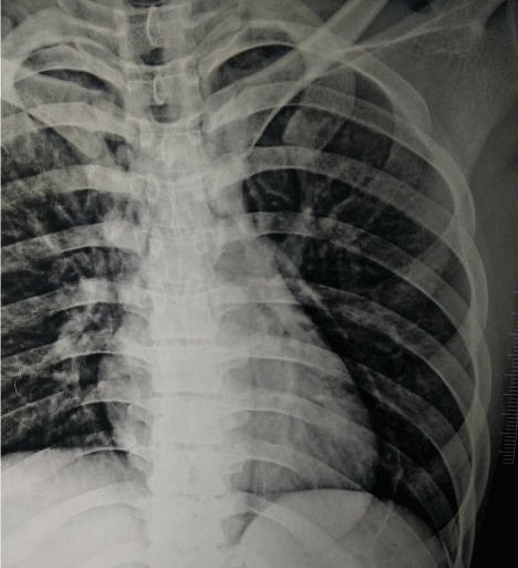
A 34-year-old man presented to the emergency room 6 hours after a road traffic accident. He was hemodynamically and neurologically stable. A chest X- ray showed the classical ‘Halo” sign, suggestive of Pneumopericardium. The patient was observed and discharged the next day (Figure 1).
What is the Macklin effect?
The mechanism responsible for pneumopericardium is the ‘Macklin effect’ – There is initially an increased pressure gradient between the alveoli and the interstitial space. Increased pressure leads to alveolar rupture, resulting in air getting through to the pericapillary interstitial pulmonary space. This space is continuous with the peribronchial and pulmonary perivascular sheaths. From here, the air tracks to the hilum of the lung and then to the mediastinum. The development of pneumothorax indicates disruption of the visceral pleura. Pneumopericardium is suggestive of pericardial tear [1]. This may remain asymptomatic or may progress to life threatening conditions like tension pneumopericardium or cardiac tamponade.

Figure 1: Chest X-ray PA view showing The “Halo Sign”.
The symptomatic patient may present with dyspnea, cyanosis, chest pain, pulsus paradoxus, bradycardia or tachycardia. On physical examination, the patient may have the classic “Beck’s triad” – hypotension, raised JVP and distant heart sounds, when complicated by cardiac tamponade. Extension of the mediastinal air to the subcutaneous tissues via the fascial planes may lead to subcutaneous emphysema. When air and fluid mix together in the pericardial sac, a tinkling sound superimposed over a succession splash is heard. This is known as a “Bruit de Moulin”, which is French for “Mill–wheel” murmur [2]. Air between the anterior parietal pericardium and the thoracic cage may also give rise to the “Hamman’s Sign” – which is a crunching sound typically heard on auscultation of the chest, but may sometimes be heard even with the unaided ear [3]. Non-specific ECG changes such as low-voltage, ST-segment changes, and T-wave inversion can be seen but are unreliable for diagnosis. On chest radiographs, a continuous rim of air outlined by a radiopaque line representing the pericardial sac, is characteristic. This is known as the “Halo sign”. This radiolucent line of air is distinctively confined to below the aortic arch. Many etiological factors have been described for pneumopericardium, including trauma, invasive procedures, barotrauma, fistulae and pericardial infections [2]. The asymptomatic stable patient requires only observation, but patients with tension pneumothorax and tamponade require urgent pericardiocentesis.
References
- Baharudin A, Sayuti RM, Shahid H. The Macklin effect--pneumomediastinum and pneumopericardium following blunt chest trauma. Med J Malaysia. 2006; 61: 371-373.
- White C, Haramati L, Chen J, Levsky J. Pnemopericardium. Cardiac Imaging, Oxford University Press. 2014; 442-444.
- Macklin CC. Transport of air along sheaths of pulmonic blood vessels from alveoli to mediastinum: clinical implication. Arch Intern Med. 1939; 64: 913-926.