Case Report
Austin J Surg. 2016; 3(1): 1080.
Pre-Operatively Diagnosed Omental Torsion and Infarction in a Child: A Rare Case Report
Tiwari C, Shah H*, Desale J, Makhija D and Jayaswal S
Department of Paediatric Surgery, TNMC & BYL Nair Hospital, India
*Corresponding author: Shah H, Department of Paediatric Surgery, TNMC & BYL Nair Hospital, Mumbai, Maharashtra, India
Received: January 18, 2016; Accepted: March 10, 2016; Published: March 14, 2016
Abstract
Omental torsion leading to infarction is a rare cause of acute abdomen and is rarely diagnosed pre-operatively. It mimics acute abdominal conditions like appendicitis, acute diverticulitis and Meckel’s diverticulum. The patient presents with right-sided abdominal pain, especially in the paediatric age group. About 0.1% of paediatric patients who undergo laparotomy for suspected appendicitis, will have omental infarction. We describe a case of an 11 year-old boy diagnosed pre-operatively as omental torsion by contrast-enhanced Computed Tomography Scan which was confirmed at laparoscopy.
Keywords: Omental torsion; Omental infarction; Whirl sign; Laparoscopy
Case Presentation
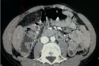
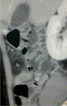
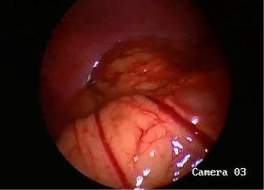
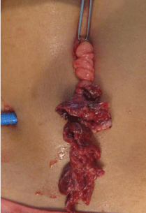
An eleven year-old boy was admitted with complaints of rightsided lower abdominal pain of one day duration. There was history of mild fever; but no history of prodromal syndromes like nausea, vomiting, anorexia, diarrhea or constipation. At admission, his vitals were stable except for tachycardia. There was severe tenderness and involuntary guarding present in right lumbar, right iliac fossa and umbilical region. Leucocytosis was present (Total Leucocyte Count: 17,600 per cu.mm). Abdominal ultrasound revealed an ill-defined area of size 5.2x1.9x4.7cm in the right para-umbilical region with hyperechoic linear strands suggesting inflamed fat abutting the anterior abdominal wall suggestive of omental torsion. Contrast Enhanced Computed Tomography (CECT) scan (Figure 1) showed a 3.7x4.8x3.4cm focal area of fat stranding in the right sub-hepatic region abutting the anterior abdominal wall inferior to the transverse colon. It contained a vascular pedicle at its centre which did not show enhancement on post contrast arterial and venous phase. Linear folds of omental tissue in concentric pattern (Whirl Sign) could be seen (Figure 2). This suggested possibility of omental torsion with thrombosed vascular pedicle resulting in secondary omental infarct. Patient was started on intravenous antibiotics and analgesics. But the pain persisted. At diagnostic laparoscopy, infarcted and necroses omentum was seen adherent to the right anterior abdominal wall near the umbilicus (Figure 2). There was minimal sero-sanguinous fluid in the peritoneal cavity. Rest of the abdomen was normal. The omentum was mobilized and delivered through the umbilical site (Figure 3). Three turns of torsion were seen and the omentum distal to it was necrosed. The omentum was excised. Patient had an uneventful recovery. Histopathology revealed necrosed omentum.

    Figure 1:  CECT scan showing a 3.7 x 4.8 x 3.4 cm focal area of fat stranding
in the right sub-hepatic region abutting the anterior abdominal wall inferior to
the transverse colon. It contains a vascular pedicle at its centre which did not
show enhancement on post contrast arterial and venous phase.

    Figure 2:  CECT image showing linear folds of omental tissue in concentric
pattern (Whirl Sign – Arrow) suggestive of Omental Torsion.

    Figure 3:  Laparoscopic image showing twisted omentum with prominent
vessels.

    Figure 4:  Twisted and necrosed omentum delivered through the umbilical
port. The omentum is twisted three turns.
Discussion
Omental torsion is a rare condition in the paediatric age group. It presents with acute right lower quadrant pain and is seen in about 0.1% of paediatric patients undergoing laparotomy for suspected acute appendicitis [1]. It was first reported by Eitel in 1899 [2,3]. It is a condition in which there is twisting of a pedicle of omentum on its longer axis to such an extent that its vascularity is compromised [2]. This leads to infarction which can range from simple edema or ischemia or even gangrene of the omentum [4].
Omental infarction usually occurs in adults in the 4th and 5th decade [1]. Males predominate over females by a ratio of 2:1 [1]. Children are less likely to get omental torsion because of less omental fat. The increasing recent reports of paediatric omental torsion can be explained by the more widespread use of CECT scan for investigating paediatric abdominal pain and the increase in incidence of paediatric obesity [4].
Omental torsion is classified as “primary” and “secondary” [5]. Primary or idiopathic torsion is seen in patients with anatomical variations such as tongue-like projections from the free edge of the omentum, bifid omentum, and accessory omentum [1,6,7]. It should be suspected in children with negative laparotomy for acute appendicitis or Meckel diverticulitis, especially in the presence of sero-sanguinous peritoneal fluid [6]. Secondary omental torsions occur due to an underlying pathology like cysts and tumours of omentum [5], bulky abdominal tumour [6], internal hernia [5,6], or as a sequela of previous abdominal surgeries [6].
Omental torsion can also be divided into “unipolar” and “bipolar.” In unipolar omental torsion, the proximal omentum remains fixed and the other tongues are free. In cases of bipolar omental torsion, both proximal and distal omenta are fixed [5]. Primary omental torsion is always unipolar, and secondary omental torsion may develop as unipolar and bipolar [5].
Predisposing factors for omental torsion include changes in omental consistency including inflammation, edema and excess fat deposition or anatomic abnormalities [5,7] and obesity. Precipitating factors include trauma, hyper-peristalsis and acute changes in body position like twisting body movements such as in wrestling and horse riding, taking strong purgative drugs, and overeating [5]. These events lead to a sudden shift of omentum, resulting in torsion [6].
The pathogenesis starts with omental congestion due to venous obstruction leading to an inflammatory response, adhesion formation, and finally, necrosis as a result of both venous and arterial obstruction [6]. This presents as abdominal pain. Spontaneous derotation of omental torsion is also possible and may explain the nonspecific omental adhesions sometimes found incidentally during laparotomy [6].
Patients present with right-flank and lower abdominal pain leading to misdiagnosis as acute appendicitis, cholecystitis, cecal diverticulitis, appendagitis, twisted ovarian cyst, etc [5,6]. The right side of the greater omentum being more mobile, omental torsion often develops on the right side. However, the prodromal symptoms like nausea, vomiting, anorexia and altered bowel habits are usually absent [1], although mild fever may be present [5].
Preoperative diagnosis of omental torsion is difficult. Abdominal ultrasound shows a wedge/triangular shaped, non-compressible hyperechoic mass deep to anterior abdominal wall [1] and free fluid within the peritoneal cavity. The sensitivity of ultrasound has been reported as 64% in the paediatric age group [1,8]. The classical signs of omental torsion on CT scan are of a hazy fatty mass with concentric linear strands in the greater omentum, the whirl sign [7,9]. These strands are twisted blood vessels whirling around a central rod [7].
The management is conservative in pre-operatively diagnosed [1] and stable patients [7]. This includes reassurance, appropriate analgesia and antibiotics. This condition is self-limiting [1] and resolution is expected in two weeks [7]. The omentum becomes atrophic and fibrotic and, on rare occasions, the pedicle may even auto-amputate (automatic clinical regression) [7]. Spontaneous derotation is also possible. If conservative management fails, treatment involves resection of the diseased segment of omentum and correction of any secondary pathology, if present. Sometimes, untreated omental torsion can lead to intra-abdominal abscesses, peritonitis, or bowel obstruction. Complications include omental necrosis, peritonitis, bowel obstruction, adhesion formation, and sepsis which make its early diagnosis critical.
References
- Robertson I, O’Connor D, Khan W, Barry K. Pediatric omental infarction: value of the laparoscope. Int J Case Rep Images. 2015; 6: 325-327.
- Ghosh Y, Arora R. Omental Torsion: A review of Literature and case report. Ind J Cli Practice. 2014; 24: 987-988.
- Eitel GG. Rare omental torsion. NY Med Rec. 1899; 55: 715-716.
- Chen CD, Chen XM, Wu SC. A child with the rare diagnosis of acute appendicitis and omental torsion. HK Paediatr (new series). 2012; 17: 254- 257.
- Katagiri H, Honjo K, Nasu M, Fujisawa M, Kojima K. Omental Infarction due to Omental Torsion. Case Rep Surg. 2013; 2013: 373810.
- Tandon AA, Lim KS. Torsion of the greater omentum: A rare preoperative diagnosis. Indian J Radiol Imaging. 2010; 20: 294-296.
- Jain P, Chhabra S, Parikh K, Vaidya A. Omental torsion. J Indian Assoc Pediatr Surg. 2008; 13: 151-152.
- Rimon A, Daneman A, Gerstle JT, Ratnapalan S. Omental infarction in children. J Pediatr. 2009; 155: 427-431.
- Lee W, Ong CL, Chong CC, Hwang WS. Omental infarction in children: imaging features with pathological correlation. Singapore Med J. 2005; 46: 328-332.