Clinical Image
Austin J Surg. 2016; 3(2): 1085.
Mediastinal Liposarcoma
Tamer Shalaby*
Department of General and Acute Medicine, Ashford and St Peters Hospitals, UK
*Corresponding author: Tamer Shalaby, Department of General and Acute Medicine, Ashford and St Peters Hospitals, Bournewood House, Guildford Rd, Chertsey KT16 0QA, UK
Received: July 04, 2016; Accepted: July 11, 2016; Published: July 20, 2016
Clinical Image
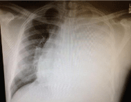
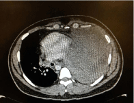
A 36 year old man, non smoker who is previously fit and well presented with 10 days history of progressive shortness of breath on exertion, dry cough and dizziness with minimal constitutional symptoms. He was in respiratory distress on examination and his chest x-ray showed complete white left lung with significant mediastinal shift (Figure 1) and an urgent bedside thoracic ultrasound showed a large area of solid mass and did not show much effusion and hence a CT chest was requested. Findings where that of missed density mass filling much of the left hemithorax and invading the mediastinum (Figure 2). The ultrasound guided biopsy result showed well-differentiated mediastinal liposarcoma.

Figure 1: Chest x-ray.

Figure 2: CT chest.
Primary mediastinall iposarcomes are extremely rare. Four main types of liposarcomas have been described: myxoid, well differentiated, and dedifferentiated and pleomorphic. Welldifferentiated low-grade liposarcomas have histologic features in many areas resembling mature adipose tissue [1]. Interspersed areas contain lipocytes that exhibit greater variation in size and shape than the lipocytes observed in lipomas. Scattered cells in these areas and in fibrous bands show atypical cytologic features [1]. On CT imaging, mediastinal liposarcomas appears as inhomogeneous fatty masses that vary in appearance depending on the amount of soft tissue and fibrous bands in the tumour. These morphologic features along with a rapid rate of growth and tracheal or vascular displacement should raise the suspicion for liposarcom [2]. The recommended treatment for mediastinal liposarcoma is surgical excision. If the entire tumour cannot be resected, surgical debulking often results in symptomatic relief. Radiation therapy and chemotherapy may be added as adjuncts to surgical excision [3].
References
- Evans HL. Liposarcomas and atypical lipomatoustumors: a study of 66 cases followed for a minimum of 10 years. SurgPathol. 1988; 1: 41-54.
- Munk PL, Lee MJ, Janzen DL, Connell DG, Logan PM, Poon PY, et al. Lipoma and liposarcoma: evaluation using CT and MR imaging. AJR. 1997; 169: 589-594.
- Grewal RG, Prager K, Austin JHM, Rotterdam H. Long term survival in nonencapsulated primary liposarcoma of the mediastinum. Thorax. 1993; 48: 1276-1277.