Special Article - Surgical Case Reports
Austin J Surg. 2016; 3(3): 1089.
Application of three-Dimensional Printed Acetabular Model to the Treatment of Severe Acetabular Bone Defect in Cup Revision Surgery: A Technical Case Report
Hoshino H*, Koyama H, Nishikino S and Matsuyama Y
Department of Orthopedic Surgery, Hamamatsu University School of Medicine, Japan
*Corresponding author: Hoshino H, Department of Orthopedic Surgery, Hamamatsu University School of Medicine, 1-20-1 Handayama, Hamamatsu, 431-3192, Japan
Received: October 04, 2016; Accepted: October 17, 2016; Published: October 18, 2016
Abstract
The application of three-dimensional (3D) printed acetabular model made by personal 3D printer to cup revision surgery has some advantages such as preoperative evaluation, shorting operation time and easier creation of fitted bulk allograft in the cases with a massive bone defect which would need a structural bulk allograft along with the Kerboull-type acetabular reinforcement device. We presented a cup revision case with severe acetabular bone defect treated by the surgical technique for the intraoperative simulation using 3D printed acetabular model to create the fitted structural bulk allograft. There were no obvious complications such as post-operative infection associated with 3D printed models although the patient was observed for a short term period. This technique using 3D printed model has the great advantages, which would be a useful option for cup revision surgery with massive defect of the acetabulum.
Keywords: 3D Printing; Hip joint; Cup revision; Acetabular model
Introduction
Acetabular cup revision surgery for the patients with a massive bone defect in the acetabulum often requires an acetabular reinforcement device with bone allograft for stable fixation of acetabular cup [1,2]. We treated Type III defects according to the American Academy of Orthopaedic Surgeons (AAOS) classification [3], with a structural bulk allograft and morsellised allograft bone chips along with the Kerboull-type acetabular reinforcement device (KT-plate; Japan Medical Materials, Osaka, Japan), which was made of titanium to support allografts and useful device for stable acetabular reconstruction [4]. However, it is not so accurate to estimate the size of the defect before operation and it is time-consuming to fit a structural bulk allograft into the shape of segmental defect between acetabular site and KT-plate during operation. It might be useful for surgeon to estimate the size of the defect directly before the operation and to create the fitted structural bulk allograft outside the body separately.
We present a cup revision case with severe acetabular bone defect treated by the surgical technique for the intraoperative simulation using three-dimensional printed acetabular model to create the fitted structural bulk allograft.
Case Presentation
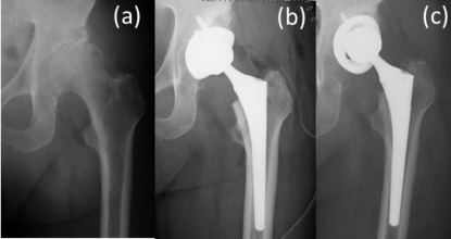
A 68-year-old man who has osteoarthritis of the right hip joint was treated by cementless total hip arthroplasty (Figure 1). The cup implant gradually loosened and migrated superiorly because of the collapse of the acetabular cyst three months after operation. During the time after operation, his left hip pain was getting worse and did not improve. Computed tomography revealed a segmental defect at the acetabulum, corresponding to AAOS type 3.

    Figure 1:  Radiograph of the patient showing, (a) osteoarthritis with a large
cyst in the left hip joint before initial operation, (b) total hip arthroplasty one
week after operation, and (c) cup implant loosened and migrated superiorly
3 months after operation.
After obtaining informed consent, the DICOM images were processing software (Mimics 3-matic, Materialise, Belgium) for creating the 3D acetabular image. The STL file of the 3D acetabular image was transferred to the personal 3D printer (UP!Plus 2, SUNSTELA, Japan) to create the 3D full-scale acetabular model made by acrylonitrile-butadiene-styrene resin. At this moment we estimated the size of the defect and the amount of allograft bone needed by the simulation of trial KT plate placement. The printed 3D full-scale acetabular model was sterilized by ethylene oxide gas with 55 degrees C for 60 minutes before the operation.
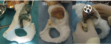
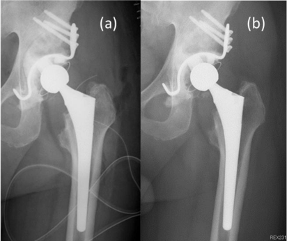
The patient was placed in a lateral decubitus position with general anesthesia. The 3D model was placed another sterilized table. The surgical approach was anterolateral mini-one incision [5]. After removal of the cup implant, acetabular bone with massive defect was exposed and prepared by removal of adhering tissues. At the same time, the creation of bulk allograft made by femoral head which was obtained from bone bank to be fitted to the curvature of the defected acetabulum and the simulation of KT plate placement with created bulk allograft using 3D printed acetabular model was performed by an assistant doctor at another sterilized table (Figure 2). After the preparation of defected acetabulum and the creation of fitted bulk bone with defected acetabulum, the morsellised bone chips which were also obtained from femoral head were impacted as firmly as possible for cavitary defect and the created bulk bone was placed for segmental defect. The hook of the KT plate was placed under the teardrop area and KT plate was fixed to the pelvic bone with three screws partly passed through the placed bulk bone. After that, a polyethylene acetabular component was fixed with cement to the KT plate (Figure 3a). The operation time was 2 hours and 18 minutes and total blood loss was 560ml. The patient started to ambulate with walker at one week after operation. The modified Harris hip score was 93 points and X-ray photograph showed no remarkable changes in the revised cup implant at 15 months after operation (Figure 3b).

    Figure 2:  Intra-operative simulation using 3D printed model showing (a)
the estimate for the size of the defect and the amount of allograft bone
needed by the simulation of trial KT plate placement, (b) the creation of bulk
allograft made by femoral head to be fitted to the curvature of the defected
acetabulum, and (c) the simulation of KT plate placement with created bulk
allograft using 3D printed acetabular model performed by an assistant doctor
at another sterilized table.

    Figure 3:  Post-operative radiograph showing (a) just after surgery, and (b)
18 months after surgery.
Discussion
Three-dimensional printing technology is being rapidly developed in the orthopedic field [6,7]. It allows surgeon not only to combine imagination with practical 3D pathologies, but also to perform surgery more accurately and handily. Especially, 3D printing technology might be useful for orthopedic surgeon to perform cup revision surgery with massive defect of accetabulum because of the anatomical complexity of pelvis. Some authors reported the usefulness of 3D printing for the treatment of pelvic fracture [8,9].
In our technique, the application of 3D printed acetabular model made by personal 3D printer to cup revision surgery has some advantages in the cases with a massive bone defect which would need a structural bulk allograft along with the Kerboull-type acetabular reinforcement device. The first is to evaluate the extent of acetabular bone defect with the simulation of KT plate placement to the 3D printed acetabular model in advance preoperatively. The second is to create a bulk allograft fitted to the curvature of the defected acetabulum of the sterilized 3D printed model at another table by another doctor in parallel with the actual operation, which would be conducive to shorting operation time and easier creation of fitted bulk allograft. The third is to create this model at a lower cost using the personal 3D printer compared with outsourcing the creation of this model to professional corporations in Japan. On the other hand, there are a couple of drawbacks such as the risk of infection and the limitation for the condition of sterilization because the created model made by acrylonitrile-butadiene-styrene resin could deform at the heat condition above 80 degrees Celsius.
In our case, there were no complications such as post-operative infection associated with 3D printed models although the patient was observed for a short term period. This technique using 3D printed model has the great advantages, which would be a useful option for cup revision surgery with massive defect of the acetabulum.
Consent
Written informed consent was obtained from the patient for publication of this case report and accompanying images.
References
- Kerboull M, Hamadouche M, Kerboull L. The Kerboull acetabular reinforcement device in major acetabular reconstructions. Clin Orthop. 2000; 378: 155-168.
- Winter E, Piert M, Volkmann R, Maurer F, Eingartner C, Weise K, et al. Allogeneic cancellous bone graft and a Burch-Schneider ring for acetabular reconstruction in revision hip arthroplasty. J Bone Joint Surg [Am]. 2001; 83- A: 862-867.
- D’Antonio JA, Capello WN, Borden LS, Bargar WL, Bierbaum BF, Boettcher WG, et al. Classification and management of acetabular abnormalities in total hip arthroplasty. Clin Orthop. 1989; 243: 126-137.
- Tanaka C, Shikata J, Ikenaga M, Takahashi M. Acetabular reconstruction using a Kerboull-type acetabular reinforcement device and hydroxyapatite granules: a 3-to 8-year follow-up study. J Arthroplasty. 2003; 18: 719-725.
- Berger RA. Mini-incision total hip replacement using an anterolateral approach: technique and results. Orthop Clin North Am. 2004; 35: 143-151.
- Jeong HS, Park KJ, Kil KM, Chong S, Eun HJ, Lee TS, et al. Minimally invasive plate osteosynthesis using 3D printing for shaft fractures of clavicles: technical note. Arch Orthop Trauma Surg. 2014; 134: 1551-1555.
- Hirano M, Ikemoto S, Tsuboi H, Akita S, Ohshima S, Saeki Y, et al. Computer assisted planning and custom-made surgical guide for malunited pronation deformity after first metatarsophalangeal joint arthrodesis in rheumatoid arthritis: a case report. Comput Aided Surg. 2014; 19: 13-19.
- Wu XB, Wang JQ, Zhao CP, Sun X, Shi Y, Zhang ZA, et al. Printed threedimensional anatomic templates for virtual preoperative planning before reconstruction of old pelvic injuries: initial results. Chin Med J. 2015; 20: 477- 482.
- Zeng C, Xiao J, Wu Z, Huang W. Evaluation of three-dimensional printing for internal fixation of unstable pelvic fracture from minimal invasive pararectus abdominis approach: a preliminary report. Int J Clin Med. 2015; 15: 13039-13044.