Case Report
Austin J Surg. 2018; 5(3): 1132.
Peripheral Desmoplastic Ameloblastoma: Report of an Extremely Rare Presentation and a Literature Review
Ramphul A¹*, Kho Y² and Mehanna P¹
¹Oral and Maxillofacial Surgery Registrar, Division of Surgery, John Hunter Hospital, Australia
²Resident Medical Officer, Prince of Wales Hospital, Australia
*Corresponding author: Ramphul A, Oral and Maxillofacial Surgery Registrar, Division of Surgery, John Hunter Hospital, Newcastle, Australia
Received: January 09, 2018; Accepted: February 12, 2018; Published: February 22, 2018
Abstract
Thedesmoplastic variant of Peripheral Ameloblastomais an extremely rareform of Ameloblastoma. There are only five reported cases of Peripheral Desmoplastic Ameloblastoma (PDA) in the English literature. Peripheral Desmoplastic Ameloblastomapresents as a soft tissue mass on the tooth bearing areas of the jaw and has very distinct histological features, characterised by de novo synthesis of collagen. In general, Peripheral (extra osseous) forms of Ameloblastomaare considered to be less aggressive their Central (intra osseous) variant, with significant difference in their management. However, the paucity of information on the much rarer desmoplastic subtypes of Peripheral Ameloblastoma makes it impossible to assess its biologic behaviour. Clinical vigilance is hence warranted with this form of Ameloblastoma. We reviewed the literature and present the case of a 60-year-old male presenting with a PDA on the mandibular attached gingival in the right first premolar area.
Introduction
Ameloblastoma is a slow growing tumour of the jaw. It is a tumour of odontogenic origin and accounts for 1% of all oral tumours and about 18% of odontogenic tumours [1]. It can present either as an intra osseous or an extra osseous lesion. The extra osseous form arises from the gingival or alveolar mucosa and is known as Peripheral Ameloblastoma (PA).
The follicular and plexiform variants are the most common histological subtypes of ameloblastoma. Less common histological variants include the acanthomatous, granular, basal cell, keratoameloblastoma and clear cell subtypes [2]. The desmoplastic subtype of Ameloblastoma is very rare. There does not appear to be a relationship between the histology of the more common histological variants and prognosis [3]. On the other hand, it is hard to predict the biological behaviour of the desmoplastic variant due to the paucity of information in the literature.
Peripheral Ameloblastoma is a soft tissue tumour occurring on the tooth bearing areas of the maxilla or mandible [4] and most commonly manifests as a slow growing, firm, painless mass with a sessile or pedunculated base. It usually has a smooth texture but can sometimes be pebbly, granular, papillary or warty [4,5] with a colour that can vary from that of normal mucosa to pink, red or dark red [5,6]. The lesion can also demonstrate an ulcerated or a keratotic appearance (frictional keratosis) due to trauma during mastication [4]. The mandibular premolar area is the most common site of presentation, accounting for 32.6% of all sites [4]. It is common for PA to erode cortical bone by mass effect. This is referred to as cupping or saucerisation.
The desmoplastic variant of Peripheral Ameloblastoma is an extremely rare form of Ameloblastoma and presents with very distinct histological features, with only 3 reported cases that we have been able to identify in the literature.
The authors describe a case of a PDA in the mandibular premolar area in a 60 year old and review the literature on the subject.
Case Presentation
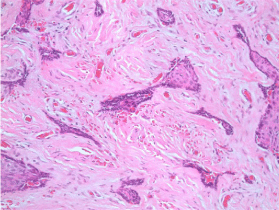
A 60-year-old man presented with a few weeks history of a hard gingival swelling on the buccal gingival of his first right mandibular premolar (44). A well-circumscribed firm lesion around 7 mm in diameter was noted in the right canine/first premolar area, with no pain or mobility of any of the adjacent teeth (Figure 1). The lesion was non-tender with no gingival erythematic, ulceration or bleeding noted or reported. Cone Beam CT showed an expansive radiolucent lesion in the premolar/canine area (Figure 2). Following discussions with the patient, he underwent an excision biopsy of the lesion. The biopsy specimen showed squamous mucosa and sub mucosa with small bone trabeculae present on the deep surface (Figures 3,4). In the sub mucosa there was a circumscribed neoplasm consisting of nests of cells in a fibrotic stroma with the nest having a peripheral layer of palisade small cuboidal cells with a high Nuclear: Cytoplasmic ratio, oval, even nuclei and scanty neoplasm. The central cells were squamoid with some squamous nests. There was no necrosis, mitosis nor were pleomorphism and the histological findings consistent with the diagnosis of Peripheral Desmoplastic Ameloblastoma (PDA). The peripheral margins of the specimen were clear but the deep margin was positive. The patient had surgery again where the deep margin was mechanically derided with a round bur by 2-3 mm. This time, the histopathology analysis reported a clear margin. The patient has now been followed up for three years and there is no evidence of recurrence.

    Figure 1:  Peripheral Desmoplastic Ameloblastoma lesion presenting as a
hard bony swelling in the canine/premolar area.

    Figure 2:  Well circumscribed expansile radioluscent lesion presenting on the
buccal cortex on mandible in the canine/premolar area.

    Figure 3:  Squamous mucosa and submucosa with bone trabeculae, nest of
cells with fibrotic stroma.

    Figure 4:  Squamous mucosa and submucosa with bone trabeculae, nest of
cells with fibrotic stroma.
Discussion
Stanley and Krogh were the first to report a case of Peripheral Ameloblastomain 1959. It is a rare extra osseous tumour that shares similar histological features to its intraosseous variant, the centrally located ameloblastoma. It accounts for 2 to 10% of all ameloblastomas [7] and is described as a benign neoplasm (or hamartomatous lesion) confined to the soft tissue overlying the tooth bearing areas of the jaw [4].
Clinical Features
In a review of 160 cases, Philipsen et al. concluded that the mean age of onset of Peripheral (extra osseous) Ameloblastoma (PA) was 52.1 years [4], which is in contrast to central (intra osseous) ameloblastoma that tends to occur at a younger age. Our patient was 60 years old. PA occurs more commonly in the mandible (70.9%) than in the maxilla (29.1%) [4]. Most of the maxillary PA reported were located in the posterior part of the hard palate or in the soft palate, in the tuberoses area [4,8] with very few reports of PA in the labial part of the maxilla [8]. The mandibular premolar area is the most common site of presentation, accounting for 32.6% of all cases [4]. The lesion in our case was located in the gingival mucosa of the teeth 43/44 area. Zhu et al, in a review of 16 cases of PA, also reported the canine premolar area in the mandible as the most common site of presentation followed by the molar and incisor region [9].
Clinical and Radiographic Presentation
Clinically, PA presents as a painless, sessile, firm and exophytic growth [4]. Its size varies from 0.3 to 4.5 cm in diameter with a mean of 1.3cm [4]. Its differential diagnosis includes fibrous epilus, Peripheral Giant Cell Granuloma, Peripheral Odontogenic Fibroma, Peripheral Ossifying Fibroma, papilloma and pyogenic granuloma [4-6]. There is usually no bony involvement on radiograph; however a superficial erosion of bone can often be noticed on radiograph or at surgery. This is known as cupping or saucerisation and is attributed to pressure resorption rather than neoplastic erosion [4].
The more common histological presentations of PA such as follicular, plexiform, basal cell and acanthomatous are usually considered to be non aggressive [4]. Gardner believes that the dense fibrous tissue of the gingival and periosteum as well as cortical plate of the alveolar process can possibly act as a barrier to bony infiltration by a PA [10].
Histology
Histological, there is no difference in cell type and pattern between the Peripheral and a Central Ameloblastoma.
The pathophysiology of PDA is not well understood. There are two proposed histological origins of PDA [11-14]. It is thought that tumours that show complete separation from overlying surface epithelium arise from odontogenic epithelial remnants [11-14]. On the other hand, the tumours that show a direct extension from the surface epithelium could arise from the basal cell layer of the overlying epithelium [11-14].
PA consists of proliferating odontogenic epithelium that exhibits the same histomorphologic cell types and patterns as seen in the intraosseous or infiltrating ameloblastoma. It can be of the follicular, plexiform, basal cell, acanthomatousor desmoplastic subtype. According to Gardner, the acanthomatous form is the most common subtype in the peripheral lesions [15]. The stroma in PA is that of a mature fibrous connective tissue. Occurrence of calcifications, dentinoid, bone like or cementum like masses is not characteristic histological features of PA [4].
Desmoplastic Ameloblastoma is a newly recognised rare form of odontogenic neoplasm [16]. It is a histological subtype of Ameloblastoma and is characterised by a hypocellular desmoplastic stroma consisting of dense collagen fibres that compress the tumour cells into islands [2]. The most important differentiating factor between the desmoplastic and other histological forms of Ameloblastoma is the abundance of thick collagen fibres in the stroma of the desmoplastic subtype [17].
In a comparative immunohistochemical study, Becker et al demonstrated that the stroma in desmoplastic ameloblastoma exhibited a strong positive reaction for collagen type VI [18,19]. This was viewed as indicating an active de novo synthesis of extracellular matrix [18]. It was hence concluded that the desmoplastic stroma did not constitute scar tissue but rather, newly produced connective tissue [18]. Due to the paucity of reports on Desmoplastic Ameloblastoma, it is difficult to judge its biologic behaviour [17]. The few reported cases are of the central intra osseous type. This case is only the 6th reported case of Peripheral Desmoplastic Ameloblastoma in the English literature.
Management
In general, the more common histological forms of PA are considered to bebenign lesions [4]. As opposed to its intraosseous counterpart which is locally aggressive and lead to bone destruction, hence requiring extensive surgical management, PA can be management in a conservative manner through supra periosteal surgical excision with adequate disease free margins [4]. Gardner is of the opinion that the excision must be down to periosteum and there is usually no need to remove bone or teeth [15]. Conservative excision of the tumour with minimal but adequate margins is the treatment of choice [18]. Smullin et al. recommends achieving a margin of 2-3 mm [2]. Recurrences are uncommon. They are usually due to incomplete removal rather than aggressiveness [4]. It is however important to follow up patients who underwent excision as there is a reported case of a benign appearing PA recurring as ameloblastic carcinoma [20]. Follow up visits should include both clinical examination with palpation of the tissues and radiographic examination.
Conclusion
The biological behaviour of Peripheral Desmoplastic Ameloblastoma is still unknown due to the paucity of reported cases. This makes it difficult to establish management guidelines. It is being managed in much more conservative manner than its central intra osseous variant. Clinicians should be encouraged to share their experience on this rare histological variant of Ameloblastoma. Until we know more about its behaviour, clinical vigilance is warranted in regards to management and follow-up.
References
- Lee SK, Kim YS. Current concepts and occurrence of epithelial odontogenic tumors: I. Ameloblastoma and adenomatoid odontogenic tumor. Korean J Pathol. 2013; 47: 191-202.
- Smullin SE, Faquin W, Susarla SM, Kaban LB. Peripheral desmoplastic ameloblastoma: report of a case and literature review. Oral Surg Oral Med Oral Pathol Oral Radiol Endod. 2008; 105: 37-40.
- Ferretti C, Polakow R, Coleman H. Recurrent ameloblastoma: report of 2 cases. J Oral Maxillofac Surg. 2000; 58: 800-804.
- Philipsen HP, Reichart PA, Nikai H, Takata T, Kudo Y. Peripheral ameloblastoma: biological profile based on 160 cases from the literature. Oral Oncol. 2001; 37: 17-27.
- Yadav RGA, Sharma R, Narain S. Peripheral Ameloblastoma: Review of the literature and case presentation. Indian Journal of Multidisciplinary Dentistry. 2011; 1.
- Reddy JKS, Kanth R, Kumar S, Reddy VK. Peripheral Ameloblastoma, a case report. Journal of Clinical and Diagnostic Research. 2011; 5: 1481-1482.
- Siar CH, Ng KH, Ngui CH, Chuah CH. Atypical peripheral ameloblastoma of the palate. J Laryngol Otol. 1990; 104: 252-254.
- Yanamoto S, YS Kawasaki G, Mizuno A. Peripheral Ameloblastoma in the maxillary Canine region. Asian J Oral Maxillofacial Surg. 2005; 17: 195-198.
- Zhu EX, Okada N, Takagi M. Peripheral ameloblastoma: case report and review of literature. J Oral Maxillofac Surg. 1995; 53: 590-594.
- DG G. Some current concepts on the pathology of ameloblastoma. Oral surgery Oral Medicine Oral Pathology Oral Radiology Endodontics. 1996; 82: 660-669.
- Vanoven BJ, Parker NP, Petruzzelli GJ. Peripheral ameloblastoma of the maxilla: a case report and literature review. Am J Otolaryngol. 2008; 29: 357- 360.
- Ide F, Mishima K, Miyazaki Y, Saito I, Kusama K. Peripheral ameloblastoma in-situ: an evidential fact of surface epithelium origin. Oral Surg Oral Med Oral Pathol Oral Radiol Endod. 2009; 108: 763-767.
- Patrikiou A, Papanicolaou S, Stylogianni E, Sotiriadou S. Peripheral ameloblastoma. Case report and review of the literature. Int J Oral Surg. 1983; 12: 51-55.
- Califano L. Peripheral ameloblastoma: report of a case with malignant aspect. Br J Oral Maxillofac Surg. 1996; 34: 240-242.
- Gardner DG. Peripheral ameloblastoma: a study of 21 cases, including 5 reported as basal cell carcinoma of the gingiva. Cancer. 1977; 39: 1625-1633.
- Eversole LR, Leider AS, Hansen LS. Ameloblastomas with pronounced desmoplasia. J Oral Maxillofac Surg. 1984; 42: 735-740.
- Philipsen HP, Reichart PA, Takata T. Desmoplastic ameloblastoma (including “hybrid” lesion of ameloblastoma). Biological profile based on 100 cases from the literature and own files. Oral Oncol. 2001; 37: 455-460.
- Buchner A, Sciubba JJ. Peripheral epithelial odontogenic tumors: A review. Oral Surgery, Oral Medicine, Oral Pathology. 1987; 63: 688-697.
- Becker J, Philipsen HP. Comparative Immunohistochemical study of the follicular and the desmoplastic ameloblastoma.
- Baden E, Doyle JL, Petriella V. Malignant transformation of peripheral ameloblastoma. Oral Surg Oral Med Oral Pathol. 1993; 75: 214-219.