Special Article – Neuro Surgery
Austin J Surg. 2018; 5(4): 1134.
Clinical Follow-Up Study for Sphera Duo® Hydrocephalus Shunt
Pinto FC, Oliveira MF*, Nespoli VS, Castro JPS, Morais JVR, Pinto FMG and Teixeira MJ
Group of Cerebral Hydrodynamics, Division of Functional Neurosurgery, University of São Paulo, Brazil
*Corresponding author: Matheus Fernandes de Oliveira, Group of Cerebral Hydrodynamics, Division of Functional Neurosurgery, Institute of Psychiatry, Hospital das Clínicas, University of São Paulo, Rua Loefgren, 700, apto 103, Vila Clementino, São Paulo, CEP 04040-000, Brazil
Received: December 19, 2017; Accepted: February 19, 2018; Published: February 26, 2018
Abstract
Cerebral hydrodynamics complications in shunted patients are due to malfunction of the system. The objective of this retrospective, single-center, single-arm cohort study is to confirm safety and performance of Sphera® Duo when used in adult patients suffering from hydrocephalus, pseudotumor cerebri or arachnoid cysts. Data were generated by reviewing 55 adult patient’s charts that were submitted to a ventriculoperitoneal shunt surgery and followed for one year after surgery. The result shows us that 85.4% of the patients improved the neurological symptoms and the reoperation rate was 12.5% in the first year after surgery.
Keywords: Ventriculoperitoneal shunt; Complications; Reoperation; Outcome
Introduction
Hydrocephalus, pseudotumor cerebri and aracnoid cysts are the main causes of cerebral hydrodynamics disturbance in adults. The surgical treatment is attained through the implantation of ventricular (to peritoneum, atrium or pleural cavity) shunt system, neuroendoscopy or both for neurological improvement [1,2].
In 1997, the United Kingdom Shunt Registry showed in 13,206 adults with hydrocephalus submitted to ventriculoperitoneal shunt (VP) implantation that 22% of all patients required reoperation within five years [3].
Cerebral hydrodynamics complications in shunted patients are due to malfunction of the system. If the shunt malfunctions and if the mechanism causing the cerebral hydrodynamics disturbance is still active, symptoms of hydrocephalus, pseudotumor cerebri or arachnoid cyst recur, and a shunt revision or other drainage procedure are required [1,2,4,5].
Malfunction may be caused by infection or mechanical failure. Approximately 40% of standard shunts malfunction occur within the first year after placement and 5% per year malfunction in subsequent years [4].
The objective of this study was to confirm safety and performance of Sphera® Duo when used in adult patients suffering from hydrocephalus, pseudotumor cerebri or arachnoid cyst.
Methods
This is a retrospective, single-center, single-arm cohort study approved by the Institutional Ethics Committee. The data are generated by reviewing 55 adult patient’s charts who were submitted to a VP shunt surgery for the treatment of cerebral hydrodynamics disturbs (hydrocephalus, pseudotumor cerebri or arachnoid cyst), from January 2015 to July 2016 at Instituto de Psiquiatria do Hospital das Cliacute;nicas da Faculdade de Medicina da Universidade de São Paulo. The SPHERA DUO® (HPBio, Brazil) shunt was used in all cases.
The SPHERA DUO® is a fixed pressure valve which works through a sequential double coil spring mechanism, seat and ruby sphere. According to the characteristic of the springs, three ranges of pressure difference ensure a flow of 21 mL/h, which corresponds to the physiological CSF production: low (3 to 7 cm H2O), medium (7 to 11 cm H2O) and high (11 to 14 cm H2O).
Primary endpoints
Frequency and severity of complications or side effects occurring in one year observation period following implantation are recorded.
Secondary endpoints
Clinical improvement after one year of shunt implantation: resolution of the intracranial hypertension syndrome (hydrocephalus, pseudotumor cerebri or arachnoid cyst) or improvement of Normal Pressure Hydrocephalus (NPH) triad (gait apraxia, memory alterations and urinary incontinence).
Study population
Inclusion criteria: Patient has received ventriculoperitoneal, ventriculoatrial or ventriculopleural shunt by implanting the SPHERA DUO® hydrocephalus shunt system.
Patient has been followed according the institutional preestablished routine in-patient and out-patient visits.
Age > 16 years old
Exclusion criteria: The patient received only the shunt (not the entire system - ventricular and peritoneal catheter) to treat a diagnosed over drainage in the previous implanted shunt of another brand.
The patient was treated by ventriculitis with Extraventricular Drainage (EVD) shortly before the implantation of the SPHERA DUO® hydrocephalus derivation system
Surgical procedure
The standard VP shunt implantation technique applied in our service is composed of cranial and abdominal approaches and is not different from the technique described by Choux et al. [4]. After initial approaches, we perform identification of peritoneum and catheterization of lateral ventricles. Simultaneously we create a subcutaneous tunnel to allow the passage of distal catheter. The whole system is attached and wounds are closed with tight suture.
Results
In the period of 1 year and 6 months (from January 2015 to July 2016), 252 surgeries were performed by the Group of Cerebral Hydrodynamics. Of these, 55 were included in this study according to the established criteria for structuring this cohort.
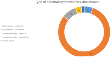
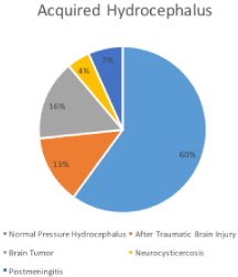
Twenty-five patients are male (45%) and 30 female (55%). The distribution of ages is represented in (Figure 1), with the youngest patient being 16 years old and the oldest being 90 years old. The most commonly treated cerebral hydrodynamic disorder is acquired hydrocephalus, accounting for 80% of cases (Figure 2), and normal pressure hydrocephalus constitutes about 60% of this sample (Figure 3). Thirty-seven valves were of medium pressure (67%), 13 of high pressure (23%) and 5 (10%) of low pressure valves were implanted according to (Table 1).
    Hydrocephalus
    Acquired
    Low pressure value
    4


    Medium pressure value
    25


    High pressure value
    8


    Congenital
    Medium pressure value
    2


    High pressure value
    2


    Pseudotumor Cerebri
    Primary
    Medium pressure value
    5


    High pressure value
    3


    Secundary
    Medium pressure value
    5


    Arcanoid Cyst

    Low pressure value
    1
Table 1: Diagnosis, classification and pressure of the valve implanted.

    Figure 1:  Distribuition of the age of the patients by range 16-25, 26-50, 51-75
and 76-90 years-old (yo).

    Figure 2:  Types of cerebral hydrodynamics disturbance: total amount
(legend) and percentage (graph).
Six patients with NPH were reoperated due to overdrainage, with replacement of medium pressure valves by high pressure ones. Overdrainage was detected in neuroimaging (tomography) examinations associated with headache and worsening of the neurological condition (memory, gait or urinary incontinence). One patient was reoperated because the distal catheter was outside the peritoneum in abdominal subcutaneous tissue.

Figure 3: Causes of the acquired hydrocephalus in the sample.
In the 12-month follow-up period, there were no cases of wound dehiscence, superficial infection or meningitis. There were no deaths related or not to surgery during the follow-up period.
Seven patients (12.5%) were reoperated in the follow-up period. One patient had to undergo the distal revision because the distal catheter migrated from the peritoneal cavity to the subcutaneous space and six patients with NPH were reoperated. Of these, five patients had the valve changed from medium to high pressure and one from low to medium pressure by hyperdrainage detected to cranial tomography by subdural collections larger than 1 cm. The latter patient has NPH, had multiple previous abdominal surgeries not related to the neurological problem, and also has pulmonary artery hypertension; having been submitted to ventricular-pleural shunt.
Patients presented radiographic improvement detected by the reduction of the Evans index, but less prominent in patients with NPH.
Forty-seven patients (85.4%) presented clinical improvement of the neurological symptoms that led to the implantation of the derivation (triad of NPH or intracranial hypertension in cases of hypertensive hydrocephalus, cerebral pseudotumor or arachnoid cyst), seven (12.7%) presented progression of NPH symptoms and one (1.9%) remained on neurological examination unchanged one year after surgery.
Discussion
Shunt infection is a common complication, occurring in approximately 5 to 15% of procedures. This may lead to ventriculitis, may promote the development of loculated compartments of Cerebrospinal Fluid (CSF), and may contribute to impaired cognitive outcome and death. The risk of shunt infections appears to be higher in newborns compared with older infants, children and adults [1-5].
Most shunt infections occur in the first six months after shunt placement. This is an important consideration in deciding when to tap shunts to evaluate a fever, especially when there is no clinical or radiographic evidence of mechanical shunt failure. Increasing abdominal pain associated with peritoneal signs and/or fever is a common presentation of shunt infection in patients with VP shunts. Abdominal ultrasound may demonstrate pseudo cyst. Shunt infection must be considered in a child with a shunt who develops persistent fever. Antibiotics should be started, but this treatment alone is often not effective. In most cases, an infected shunt must be removed, and an external ventricular drain must temporarily be placed [4-8].
Perioperative antibiotic prophylaxis reduces the risk of infection. In two meta-analyses, prophylactic antibiotics in the perioperative period reduced the risk of shunt infection by approximately 50%. The use of antibiotic-impregnated catheters also appears to lower the risk of infection. Whether prophylactic antibiotics are beneficial after the perioperative period remains uncertain. In our study we didn’t have any case of infection probably because we adopt a strict protocol using perioperative antibiotics, two gloves and the same staff always perform de VP shunt implantation [4,5].
Mechanical shunt failure is another important cause of shunt failure. Like shunt infection, it is most common during the first year after shunt placement. The majority of shunt failures result from obstruction at the ventricular catheter. Fractured tubing is the cause of shunt failure in approximately 15% of cases. Other causes include shunt migration (partial or complete) and excessive CSF drainage (over drainage). Mechanical failure requires prompt recognition and surgical intervention [2,8-11].
Over drainage can cause functional shunt failure, which causes subnormal ICP (particularly in the upright position) and which is associated with characteristic neurological symptoms such as postural headache and nausea. Over drainage greatly reduces the size of the ventricles causing the catheter to lie against the ependyma and choroid plexus, and these tissues block the holes in the catheter [8-11]. Over drainage can lead to slit-ventricle syndrome, which is characterized by small or slit-like ventricles, coupled with transient episodes of symptoms of raised ICP. Changes in shunt design to address the problem of over drainage include valves designed to open at different pressures and selected based upon the patient’s characteristics; anti-siphoning devices to minimize the siphon effect caused by changes in posture; and valves that regulate by flow rather than by pressure differences [8-11].
Six of 27 NPH patients (22.2%) in this study presented over drainage after shunting. They were submitted to reoperation. The pressure of the valve was changed medium to high in 5 cases and low to medium in one. All of them recover the neurological status prior to over drainage. The reoperation could be avoided with implantation of programmable valve or with antisifon device, but none of them developed subdural hematoma [6-8].
Other less common complications are related to the end site of CSF drainage. Potential complications in patients with VP shunts include perforation of viscus and intestinal obstruction. Patients with VA shunts may develop thrombosis associated with the atrial catheter, cor pulmonale, or very rarely may develop glomerulonephritis (“shunt nephritis”), which is related to chronic infection. Patients with ventriculopleural shunts may develop pleural effusions which occasionally produce symptoms9. One case in this study needed distal shunt revision because the distal cateter went out from the peritoneal cavity to subcutaneous space. After reoperation the patient recover the neurological status. This problem could be avoided with appropriate surgical technique. A thigh suture in reto abdominal muscle aponeurosis is indicated in obese patients.
The routine performance of a brain computed tomography (CT scan) in follow-up is of undetermined clinical utility. While ventricular size may decrease postoperatively, studies have mixed results in associating this with postoperative improvement. Thus, CT scan cannot be considered a reliable indicator of shunt functioning. CT scan may also detect a subclinical subdural effusion or hematoma.
Regular follow-up and attention to symptoms is required. When patients experience neurologic deterioration, a brain CT scan should be performed to exclude the possibility of subdural hematoma and check the catheter position. A shunt series of a plain x-ray films that visualize the entire shunt system should be performed, looking for visible obstruction. An abdominal ultrasound may also detect obstruction of the shunt tip [10,11].
Conclusion
Sphera Duo® shunt system is safe when used in adult patients suffering from hydrocephalus, pseudotumor cerebri or arachnoids cyst. 85.4% of the patients improved the neurological symptons and the reoperation rate was 12.5% in the first year after surgery.
Conflicts of Interest
Authors declare no additional conflicts of interest. All Sphera Duo® valves used in this study were provided by Hp Bio Company according to respective material acquisition policies in Public Health System in Brazil.
References
- Oliveira MF, Pinto FC, Nishikuni K, Botelho RV, Lima AM, Rotta JM. Revisiting hydrocephalus as a model to study brain resilience. Front Hum Neurosci. 2011; 5: 181.
- Pinto FC, Saad F, Oliveira MF, Pereira RM, Miranda FL, Tomai JB, et al. Role of endoscopic third ventriculostomy and ventriculoperitoneal shunt in idiopathic normal pressure hydrocephalus: preliminary results of a randomized clinical trial. Neurosurgery. 2013; 72: 845-853.
- O’Kane MC, Richards H, Winfield P, Pickrd JD. The United Kingdom Shunt Registry. Eur J Pediatr Surg. 1997; 7: S56.
- Choux M, Genitori L, Lang D, Lena G. Shunt implantation: reducing the incidence of shunt infection. J Neurosurg, 1992; 77: 875-880.
- Camacho EF, Boszczowski I, Freire MP, Pinto FC, Guimaraes T, Teixeira MJ, et al. Impact of an educational intervention implanted in a neurological intensive care unit on rates of infection related to external ventricular drains. PLoS One. 2013; 8: e50708.
- Pinto FC, Pereira RM, Saad F, Teixeira MJ. Performance of fixed-pressure valve with antisiphon device SPHERA(®) in hydrocephalus treatment and overdrainage prevention. Arq Neuropsiquiatr. 2012; 70: 704-709.
- Oliveira MF, Saad F, Reis RC, Rotta JM, Pinto FC. Programmable valve represents an ef cient and safe tool in the treatment of idiopathic normalpressure hydrocephalus patients. Arq Neuropsiquiatr. 2013; 71: 229-236.
- Pereira RM, Suguimoto MT, Oliveira MF, Tornai JB, Amaral RA, Teixeira MJ, et al. Performance of the fixed pressure valve with antisiphon device SPHERA® in the treatment of normal pressure hydrocephalus and prevention of overdrainage. Arq Neuropsiquiatr. 2016; 74: 55-61.
- Haridas A, Tomita T, Patterson M, Armsby C. “Hydrocephalus in children: Management and prognosis.” 2017.
- Graff-Radford, DeKosky NRST, Eichler AF. “Normal pressure hydrocephalus.” 2016.
- Pinto FC, de Oliveira MF. Laparoscopy for ventriculoperitoneal shunts implantation and revision surgery. World J Gastrointest Endosc. 2014; 6: 415-418.