
Special Article – Surgery Case Reports
Austin J Surg. 2018; 5(4): 1135.
Lethal Surgical Outcome of a Large Pulmonary AVM with Severe Right to Left Shunt
Wu MH* and Wu HY
Department of Surgery, Tainan Municipal Hospital, Taiwan
*Corresponding author: Ming-Ho Wu, Department of Surgery, Tainan Municipal Hospital, 670 Chung-Te Rd, Tainan, 701 ROC, Taiwan
Received: January 02, 2018; Accepted: February 19, 2018; Published: February 26, 2018
Abstract
Between October, 2001 and January, 2007, three female patients underwent lung resection for pulmonary Arteriovenous Malformation (AVM). The last two patients both with pulmonary AVM in right middle lobe were uneventful after lobectomy. The first one patient with a large pulmonary AVM presenting severe right to left shunt died of congestive heart failure on post-operative day 12. Here, we present the peri-operative images and clinical course focusing on the case of mortality. Surgical resection of a large pulmonary AVM with severe right to left shunt should be carefully concerned.
Keywords: Pulmonary AVM; Lung resection; Right to left shunt
Introduction
Between October, 2001 and January, 2007, three female patients underwent lung resection for pulmonary arteriovenous malformation (AVM) (Table 1). The last two patients both with pulmonary AVM in right middle lobe were uneventful after lobectomy. The first one patient with a large pulmonary AVM presenting severe right to left shunt died of congestive heart failure on post-operative day 12. The event occurred 16 years ago. To share the painful experience, we herein present the unique case of mortality.
    Case
    Gender, age
    Date of operation
    Location of AVM
    operation
    Outcome


    1
    Female, 37
    Oct.18,2001
    Right lower lobe
    Arterial ligation and resection of sac
    Dead


    2
    Female, 64
    Mar.25,2002
    Right middle lobe
    lobectomy
    well


    3
    Female, 61
    Jan.2,2007
    Right middle lobe
    lobectomy
    well
Table 1: Three patients with pulmonary AVM.
Case Presentation
Presentation of the fatal case
A 37 year-old female patient presented to the out-patient-clinic on October 12, 2001 with pale in looking, dyspnea, and leg edema. Physically, bruit sounds were audible at right 4th- 5th intercostals space. She was treated as pulmonary tuberculosis 10 years before. Since the early of 2001, she persisted short of breaths and severe cough. She was admitted on October 17, 2001 for surgical intervention after confirmation of pulmonary AVM. Preoperative work-up included chest film (Figure 1), chest computed tomography (Figures 2 & 3), MRI (Figure 4), and MRA, etc. Right thoracotomy with ligation of feeding artery (Figure 5) of AVM and wedge resection of right lower lobe lung, including the sac of AVM (Figure 6) was performed on October 18, 2001. Central venous pressure was 17~19 cm H2O on postoperative day 1. Oliguria was observed on post-operative day 2. Central venous pressure was elevated to 24~29 cm H2O, and cardiac echography revealed dilated right atrium and right ventricle, and pulmonary arterial pressure 50-60 mmHg on postoperative day 6. She died of heart failure on post-operative day 12.
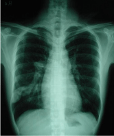
Figure 1: Chest film showed an AVM in right lung.
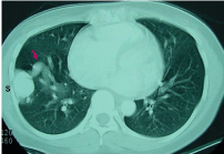
    Figure 2:  CT revealed a branch of right pulmonary artery (arrow) connecting
to the AVM (S).
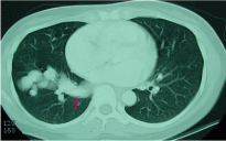
Figure 3: CT revealed predominate right inferior pulmonary vein (arrow).
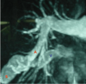
    Figure 4:  MRI revealed an AVM (S) supplied by a branch of right pulmonary
artery (A) and drained to right inferior pulmonary vein (V).
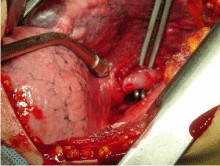
Figure 5: A sac of AVM was at the surface of right lower lobe lung.
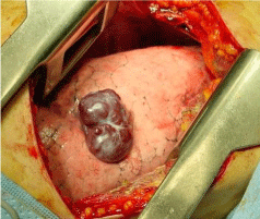
Figure 6: The feeding artery of the AVM was identified.
Comments
Pulmonary AVM have a prevalence of 1 in 2,600. Paradoxical emboli lead to strokes and cerebral abscesses [1]. Untreated pulmonary AVM had risk of stroke, brain abscess and high mortality rate [2]. The Mayo Clinic experience suggested a morbidity of 26-33% and mortality of 8-16% in untreated patients [3]. Complications of the pulmonary AVM also included intrapleural rupture and massive hemoptysis [4]. The high incidence of associated major neurologic complications mandates aggressive treatment whenever feasible. Two therapeutic options are transcatheter embolization and surgical resection [5]. Pulmonary AVM was historically treated with surgical resection [6]. Balloon embolization offers an alternative therapy for patients who are poor surgical risks or those whose lesions are too numerous to resect [5]. Transcatheter embolization is less invasive procedure with success rate of 85-98% [6]. Most surgical resections were performed using thoracotomy [7,8]. Recently, thoracoscopic surgical resection for well-selected patients with pulmonary AVM was recommended [9]. The minimally invasive lung resection was also suggested for the treatment of solitary large arteriovenous malformations [10]. Pulmonary AVM with severe hypoxemia could be considered for lung transplantation [11]. In our fatal case was treated as pulmonary tuberculosis in other hospital 10 years before. The pulmonary AVM was not recognized at that time. The large AVM with severe right to left shunt leaded to poor outcome. Lung transplantation was mentioned by a cardiologist on postoperative day 4. Majumdar etc. reported a salvage pneumonectomy for a 12-yearold boy with a large right to left shunt pulmonary AVM resulted in fatal outcome. Comparing to our presenting fatal case, careful preoperative evaluation and patient selection are necessary.
References
- Shovlin CL. Pulmonary arteriovenous malformation. Am J Respir Crit Care Med. 2014; 190: 1217-1228.
- Gossage J R, Kanj G. Pulmonary arteriovenous malformations. A state of the art review. Am J Respir Crit Care Med.1998; 158: 643-661.
- Swanson KL, Prakash UBS, Stanson AW. Pulmonary arteriovenous fistulas: Mayo Clinic experience, 1982-1997. Mayo Clin Proc. 1999; 74: 671-680.
- Thung KH, Sihoe AD, Wan IY, Lee TW, Wong R, Yim AP. Hemoptysis from an unusual pulmonary arteriovenous malformation. Ann Thorac Surg. 2003; 76: 1730-1733.
- Puskas JD, Allen MS, Moncure AC, Wain JC Jr, Hilgenberg AD, Wright C, et al. Pulmonary arteriovenous malformations: therapeutic options. Ann Thorac Surg. 1993; 56: 253-257.
- Meek ME, Meek JC, Beheshti MV. Management of Pulmonary Arteriovenous Malformations. Semin Intervent Radiol. 2011; 28: 24-31.
- Kuhajda I, Milosevic M, Ilincic D, Kuhajda D, Pekovic S, Tsirgogianni K, et al. Pulmonary arteriovenous malformation-etiology, clinical four case presentations and review of the literature. Ann Transl Med. 2015; 3: 171.
- Georghiou GP, Berman M, Vidne BA, Saute M. Pulmonary arteriovenous malformation treated by lobectomy. Eur J Cardiothorac Surg. 2003; 24: 328- 330.
- Ishikawa Y, Yamanaka K, Nishii T, Fujii K Rino Y, Maehara T. Video-assisted thoracoscopic surgery for pulmonary arteriovenous malformations: report of five cases Gen Thorac Cardiovasc Surg. 2008; 56: 187-190.
- Reichert M, Kerber S, Alkoudmani I , Bodner J. Management of a solitary pulmonary arteriovenous malformation by video-assisted thoracoscopic surgery and anatomic lingula resection: video and review. Surg Endosc. 2016; 30: 1667-1669.
- Majumdar G, Agarwal SK, Pande S, Chandra B. Salvage pneumonectomy for pulmonary arteriovenous malformation in a 12-year-old boy with brain abscess and hemiparesis: A fatal outcome. Lung India. 2016; 33: 202-204.
- Shovlin CL, Buscarini E, Hughes JMB, Allison DJ, Jackson JE. Long-term outcomes of patients with pulmonary arteriovenous malformations considered for lung transplantation, compared with similarly hypoxaemic cohorts. BMJ Open Respir Res. 2017; 13; 4.