
Special Article – Surgery Case Reports
Austin J Surg. 2018; 5(4): 1136.
Aggressive Variant of Central Giant Cell Granuloma
Ustaad F¹*, Saleem Bhat MY², Abouzeid HL³, Ali FM4 and Patil K5
1Oral & Maxillofacial Surgery, M.A Rangoonwala Dental College, India
2Department of Periodontics, Community Dental Sciences College of Dentistry, King Khalid University, Saudi Arabia
3Department of Lecturer SDS, College of Dentistry, King Khalid University, Saudi Arabia
4Department of Dentistry, Jazan University, Saudi Arabia
5Department of Oral Pathology & Microbiology, SMBT Dental College & Hospital, India
*Corresponding author: Farheen Ustaad, Oral & Maxillofacial Surgery, M.A Rangoonwala Dental College, Pune, India
Received: January 02, 2018; Accepted: February 20, 2018; Published: February 27, 2018
Abstract
Central Giant Cell Granuloma (CGCG) is relatively an uncommon central bony lesion in the head and neck region, commonly affecting the jaw bones of maxilla and mandible and has a female predilection. The clinical feature of CGCG ranges from a slow growing asymptomatic swelling to a rapidly enlarging aggressive lesion. The present case report illustrates a rare aggressive central giant cell granuloma leading to the continuity defect in the mandible and required en-bloc resection and reconstruction.
Keywords: Aggressive lesion; Central giant cell granuloma; Jaw lesions
Introduction
World Health Organization has defined Central giant cell granuloma as a “localized benign but sometimes aggressive osteolytic proliferation consisting of fibrous tissue with hemorrhage and hemosiderin deposits, presence of osteoclast-like giant cells and reactive bone formation” [1]. The clinical behavior of CGCG varies from a slowly enlarging asymptomatic swelling to an rapidly enlarging aggressive lesion that manifesting with pain, cortical perforation, and root resorption [2,3].
The pathogenesis of the CGCG of oral region has not been clearly established till date. But it is suggested as the result of an exacerbated reactive process related to previous history of trauma and hemorrhage intraosseously triggering the reparative granulomatous process. Understanding features of these benign tumors is important because they clinically mimic a malignant lesion [2-4]. The present report illustrates a rare aggressive variety of CGCG, with a typical clinical presentation in a 12-year-old female patient.
Case Report
A 12 year-old female patient reported with a complaint of swelling in the right side of body of mandible since one year. The swelling had remained asymptomatic, but gradually enlarging to cause the facial disfigurement. Neither history of trauma nor any systemic or local infections was found. The prenatal history also not found significant.
The patient presented with facial asymmetry with a poorly defined solitary swelling measuring 3 x 4 cm (Figure 1). The overlying skin appeared normal. Intraoral examination showed a diffuse enlargement of body of mandible from 43 to 45 regions, with buccal vestibule obliteration in the same region. Tooth displacement and mobility was seen with 44 and 45 but the pulp was vital with open apex. The overlying mucosa was found to be normal.
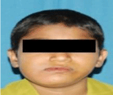
Figure 1: Extra oral swelling evident on the right mandibular region.
The swelling was bony hard, non-tender with some areas of fluctuant mass. The adjacent oral mucosa and dentition not showed any abnormality. Vitality test of associated teeth revealed normal pulpal response. A provisional diagnosis of dentigerous cyst was considered with the differential diagnosis of CGCG, ameloblastoma, odontogenic myxoma and fibrous dysplasia. OPG (Figure 2) view reveals a solitary well defined radiolucent lesion measuring 3X4 and extending to the lower border of right mandible with sclerotic border. The mandibular canal was obscured by the lesion. On aspiration the lesion had shown frank blood. After that incisional biopsy performed under local anesthesia and it showed histopathological picture composed of many multinucleated giant cells. Thus a diagnosis of CGCG was made. Laboratory values for alkaline phosphate, serum calcium, phosphorous and PTH were found to be within normal limits, as were the blood cell count, thus excluding the brown tumor of the hyperparathyroidism. The other differential diagnosis includes cherubism and aneurysmal bone cyst. Cherubism has tendency to involve bilaterally, generally occurs between the ages of 2 and 4 and often has a hereditary pattern. The two lesions giant cell tumor and CGCG are histologically almost similar, but can be differentiated from cytological features; with giant cells of the GCT presented more nuclei than those of the CGCG.
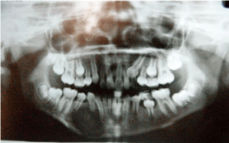
    Figure 2:  OPG showing solitary radiolucent lesion in relation to 43, 44, 45
with displacement of teeth and sclerotic borders.
On surgical exposure the lesion showed large bony expansion (Figure 3) defect involving both the buccal and the lingual cortical plate (Figure 4). Later reconstruction with an iliac graft was done. The lesion was removed (Figure 5) and the specimen was sent for the histopathology, which showed fibrocellular connective tissue stroma with numerous multinucleated giant cells (Figure 6).
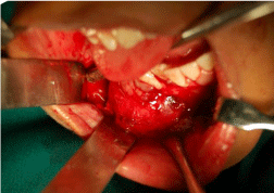
Figure 3: Bony expansion of buccal cortical plate.
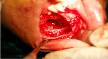
Figure 4: Perforation of the buccal and lingual cortical plate.
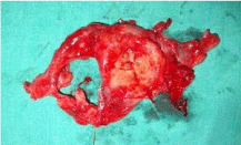
Figure 5: Photograph of the removed lesional specimen.
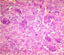
    Figure 6:  Histopathological picture of the lesion showing numerous
multinucleated giant cells with background of fibrovascular stroma.
Discussion
Jaffe in 1953 first described CGCG and it shows proliferation of multinucleated giant cells and fibroblasts [3]. CGCG is an uncommon non-odontogenic tumor and its incidence is less than 7% of all benign facial bone lesions [5,6].
CGCG is often found in the mandible, anterior to the first molars [7]. Although CGCGs are benign lesions, some authors divide CGCG into two types depending on its clinical and radio graphical features:
(a) Nonaggressive lesion- Usually asymptomatic and slow growing, does not show root perforation in teeth affected or resorption of cortical plate by the lesion, and it shows less recurrence than the aggressive type [8].
(b) Aggressive lesions- More commonly found in younger individuals and is usually painful, rapidly growing, larger in sizel, often causes root resorption and cortical perforation and also has more recurrence rate [9]. High recurrence rate of between 37.5% and 70% was found in the previous literature [7].
Radiographic appearance of CGCG can be multilocular or unilocular, with either ill-defined or well-defined margins. Tooth displacement and root resorption can be also be evident [6,10]. This case presented had a unilocular lesion with distinct and sclerotic borders. Tooth displacement was evident.
CGCG is composed of mainly of two populations of cells, multinucleated giant cells and spindle shaped stromal cells. The spindle cells are thought to be proliferating tumor cells and these are osteoblast like cells with similar functions [2,3,11].
Histologically also shows presence of hemorrhagic foci with hemosiderin pigments and occasional areas of osteoid tissue [12].
In the jaws CGCG arise either peripherally in the periodontal ligament, in the mucoperiosteum of the alveolar ridge, or centrally within the bone. Trauma is thought to be an important etiological factor. The lesion increases by accumulation of tissue, which is produced by slow, minute, continuous, multicentric haemorrhages caused by trauma and a defect in the capillaries. It has also been suggested that it could be a reaction to some form of hemodynamic disturbance in the bone marrow, perhaps associated with trauma and haemorrhage [13]. Recently, the role of c-Src was investigated in the formation of multinucleated cells in CGCG through the RANK signaling pathway [14]. Central giant cell granuloma is considered a non-reparative lesion that destroys and grows if it is not treated. Surgery is the most commonly accepted treatment modality for CGCG. But during surgery, the extent of tissue removal may be from curettage to en-bloc resection of the lesional part. Nonsurgical management is to avoid disfigurement have also been used, which includes daily doses of calcitonin and injections intralesionally of corticosteroids [15]. Osteoprotegrin, has action of inhibiting the osteoclastic bone resorption, has also been used in the treatment of CGCG. In recent years, surgical and medical combination treatment protocol consisting of curettage with removal of the peripheral bony margins and preservation of vital structures has been introduced. Also corticosteroids can be used intralesionally as a non-surgical method of management [16]. Nonsurgical treatment of CGCG is a valued management option for slowly enlarging small lesions and successful treatment of painful, rapidly enlarging large lesions is more likely to be achieved by surgical removal. In this case due to the aggressive and greater damage, surgery was needed and an en-bloc resection and reconstruction with an iliac graft was done. Although benign, damage can cause large bony defects that could lead to the Segmental resection of the mandible which causes significant patient morbidity in respect to function and esthetics. This patient follow-up was done for next 2 years and no recurrence of the lesion was evident [4,11,17].
Conclusion
The correct diagnosis of CGCG is established by correlating clinical and histological features, as CGCG in the head and neck region sometimes shows an aggressive behavior. Surgery is the commonly done and accepted treatment for large aggressive lesions which may require en-bloc resection and reconstruction which can cause significant patient morbidity.
References
- Barnes L, Eveson JW, Reichart P, Sidransky D. Pathology and Genetics of Head and Neck Tumours. Kleihues P, Sobin LH, series eds. World Health Organization Classification of Tumours. Lyon, France: IARC Press. 2005; 324.
- Ustundag E, Iseri M, Keskin G, Muezzinoglu B. Central giant cell granuloma. Int J Pediatr Otorhinolaryngol. 2002; 65: 143-146.
- Jaffe HL. Giant cell reparative granuloma, traumatic bone cyst and fibrous (fibro-osseous) dysplasia of the jawbones. Oral Surg Oral Med Oral Pathol. 1953; 6: 159-175.
- Hongal BP, Joshi P, Kulkarni V, Baldawa P. “Central giant cell granulomas of the jaws: A review of literature with its emphasis on differential diagnosis on related lesions.” Int J Contemp Dent Med Rev. 2015.
- Kaffe I, Ardekian L, Taicher S, Littner MM, Buchner A. Radiologic features of central giant cell granuloma of the jaws. Oral Surg Oral Med Oral Pathol Oral Radiol Endod. 1996; 81: 720-726.
- Sapra G, Astekar M, Hiremath SKS, Sharma M, Agrawal A, Roy S. Central giant cell granuloma of mandible. J Dent Oral Rehab. 2014; 5: 44-47.
- Rachmiel A, Emodi O, Sabo E, Azenbud D, Peled M. Combined treatment of aggressive central giant cell granuloma in the lower jaw. J Cranio-MaxilloFac Surg. 2012; 40: 292-297.
- Eisenbud L, Stern M, Rothberg M, Sachs SA. Central giant cell granuloma of the jaws: experiences in the management of 37 cases. J Oral Maxillofac Surg. 1988; 46: 376-384.
- Chuong R, Kaban LB, Kozakewich H, Perez-Atayde A. Central giant cell lesions of the jaws: a clinicopathologic study. J Oral Maxillofac Surg. 1986; 44: 708-713.
- Gungormus M, Akgul HM. Central giant cell granuloma of the jaws: A clinical and radiologic study. J Contemp Dent Pract. 2003; 4: 87-97.
- Gerzanic L, Schultes G, Kärcher H, Zebedin D. Central Giant Cell Granuloma Resistant to Calcitonin Nasal Spray: A Case Report. Anaplastology. 2013; 2: 119.
- Munzenmayer J, Tapia P, Zeballos J, Compan A, Urra A, Spencer ML. Central giant cell granuloma of the mandibular condyle. Case-report. Rev Clin Periodoncia Implantol Rehabil Oral. 2013; 6: 83-86.
- Reddy V, Saxena S, Aggarwal P, Sharma P. Incidence of central giant cell granuloma of the jaws with clinical and histological confirmation: an archival study in Northern India. Bri J Oral Maxillofac Surg. 2012; 50: 668-672.
- Wang C, Song Y, Peng B, Fan M, Li J, Zhu S, et al. Expression of c-Src and comparison of cytologic features in cherubism, central giant cell granuloma and giant cell tumors. Oncology Reports. 2006; 15: 589-594.
- Bataineh AB, Al-Khateeb T, Rawashdeh MA. The surgical treatment of central giant cell granuloma of the mandible. J Oral Maxillofac Surg. 2002; 60: 756-761.
- Ashok kumar KR, Gouder G. Central giant cell granuloma- a case report. Journal of Dental Sciences & Research. 2010; 1: 1-5.
- Gulati D, Bansal V, Dubey P, Pandey S, Agrawal A. Central Giant Cell Granuloma of Posterior Maxilla: First Expression of Primary Hyperparathyroidism. Case Reports in Endocrinology. 2015; ID 170412: 1-7.