Special Article – Colorectal Surgery
Austin J Surg. 2018; 5(5): 1139.
Right Pneumothorax Secondary to Colonoscopy-Induced Perforation
Cadili A*
Department of Surgery, University of Saskatchewan, Canada
*Corresponding author: Ali Cadili, Department of Surgery, Saskatoon, Saskatchewan, University of Saskatchewan, Canada
Received: February 26, 2018; Accepted: March 05, 2018; Published: March 12, 2018
Clinical Image
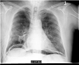
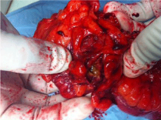
An 80 year old male presented to the Emergency Department (ED) 4 days after a screening colonoscopy with increasing shortness of breath. Chest radiography showed air under the diaphragm as well as a right pneumothorax (Figure 1). Upon review of the colonoscopy report, it was revealed that three polyps were removed from the ascending colon. The patient was resuscitated in the, placed on intravenous antibiotics, and right sided tube thoracostomy was inserted in the ED. The patient was given a provisional diagnosis of colonoscopy-induced right colonic perforation resulting in free intraperitoneal air as well as right pneumothorax. Following resuscitation, the patient was taken to the operating room on an emergency basis after a thorough discussion of the situation with the patient and his family. On laparotomy, a frank perforation was identified in the cecum with minimal gross contamination (Figure 2). The patient was also noted to be hemodynamically stable and normothermic up to that point. Ileocecal resection was performed and a primary stapled ileocolonic anastomosis was fashioned. The patient recovered well postoperatively and was discharged to a sub acute care facility for continued rehabilitation.

    Figure 1:  Chest radiograph demonstrating a right pneumothorax and free air
under the diaphragm secondary to cecal perforation.
