Special Article – Gallbladder Surgery
Austin J Surg. 2018; 5(5): 1140.
Gallbladder Radiography Following Ercp
Cadili A*
Department of Surgery, University of Saskatchewan, Canada
*Corresponding author: Ali Cadili, Department of Surgery, Saskatoon, Saskatchewan, University of Saskatchewan, Canada
Received: February 26, 2018; Accepted: March 05, 2018; Published: March 12, 2018
Clinical Image
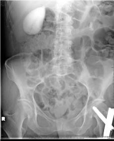
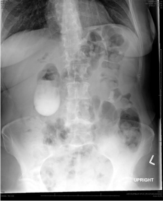
A 65 year old female was seen for new onset right sided abdominal pain 6 days post femur fracture repair. The patient’s WBC and total bilirubin were normal but ALP, ALT, and GGT were elevated. An ultrasound of the abdomen revealed a dilated common bile duct as well as intrahepatic biliary ducts with no evidence of cholecystitis or cholelithiasis. Endoscopic Retrograde Cholangio-Pancreatography (ERCP) was then performed showing no obstructive biliary lesion or stone; contrast was seen flowing freely from the cystic duct into the gallbladder during the ERCP further ruling out acute cholecystitis. Four hours after the ERCP, the patient started complaining of generalized vague abdominal pain. Abdominal and chest radiographs were obtained to rule out ERCP-induced intestinal perforation. The radiographs did not reveal any perforation but demonstrated a radiopaque gallbladder. Panels A and B show plain abdominal radiographs demonstrating a gallbladder full of residual contrast from the ERCP performed earlier (Figures 1 & 2).

    Figure 1:  It shows plain abdominal radiographs demonstrating a gallbladder
full of residual contrast.
