
Special Article – Minimally Invasive Surgery
Austin J Surg. 2018; 5(7): 1150.
Simultaneous Minimally Invasive Surgery in a Patient with Lung Cancer and Coronary Artery Disease
Kim WJ, Kang CH*, Hwang HY and Kim K-B
Department of Thoracic and Cardiovascular Surgery, Seoul National University Hospital, Korea
*Corresponding author: Kang CH, Department of Thoracic and Cardiovascular Surgery, Seoul National University Hospital, Seoul National University College of Medicine, 101 Daehak-ro, Jongno-gu, Seoul 03080, Korea
Received: September 20, 2018; Accepted: October 16, 2018; Published: October 23, 2018
Abstract
A 77-year-old woman with aggravating chest pain for a month visited our outpatient clinic. Nearly occluded left anterior descending coronary artery and right lower lobe nodule suggesting lung cancer were identified in coronary angiogram and computed tomogram, respectively. To minimize postoperative complication, simultaneous robot-assisted minimally invasive coronary artery bypass grafting (CABG) and video-assisted thoracoscopic surgery (VATS) of right lower lobectomy were performed. Postoperative course was uneventful except transient atrial fibrillation. The patient was discharged on the 8th postoperative day. Simultaneous minimally invasive CABG and VATS lobectomy instead of median sternotomy and thoracotomy approach could be a safer treatment option for concurrent coronary artery disease and lung cancer in high-risk patients.
Keywords: Minimally invasive cardiac surgery; Video-assisted thoracoscopic surgery; Coronary artery bypass graft surgery; Robot assisted surgery; Lung cancer
Case Report
A 77 years old woman visited our outpatient clinic with aggravated chest pain 1 month prior to visit. The patient underwent imaging and laboratory evaluation for possible coronary artery disease. Chest x-ray scan showed a suspicious nodular opacity in the right lower lung field. Computed tomography (CT) coronary angiography showed 90% stenosis of proximal left anterior descending coronary artery (LAD) and 1.3cm-sized part solid nodule in the right lower lobe (Figure 1). A suspicious lung cancer with a clinical stage of cT1bN0M0 was diagnosed based on the chest CT scan, positron emission tomography and bronchoscopic evaluation. Coronary angiography was performed and near total occlusion of the proximal LAD was confirmed (Figure 1). To minimize the risk of combined right lower lobectomy and coronary artery bypass grafting (CABG) in this elderly woman, simultaneous robot-assisted minimally invasive direct CABG (MIDCAB) and video-assisted thoracoscopic surgery (VATS) for right lower lobectomy was planned. The patient was positioned supine, and a camera port and 2 robot arm ports were made on the 4th and 2nd and 6th left intercostal spaces, respectively. The left internal thoracic artery (LITA) was harvested as skeletonized fashion using da Vinci Surgical System (Intuitive Surgical, Inc. Sunnyvale, CA, USA). Pericardiotomy and identification of the target vessel was followed. Then, left arm port incision was extended medially about 5cm along the sub-mammary crease to perform hand-sewn anastomosis of LITA to LAD. One more port incision was made on the 6th intercostal space to insert a shaft for a stabilizer designed for MIDCAB. After finishing robot assisted MIDCAB, the patient’s position was changed to left lateral decubitus. One utility incision was made on the 5th intercostal space along the anterior axillary line, and the other 3 additional ports were made in mid- and posterior axillary line. After confirming adenocarcinoma by intraoperative pathologic examination of frozen sectioned wedge-resected specimen, VATS right lower lobectomy and mediastinal lymph node dissection was performed. Total operation time was 330 minutes including 210 minutes for robot-assisted- MIDCAB and 120 minutes for VATS. The amount of blood loss was 80cc and no transfusion was performed. Pathologic staging was pT1aN0 with 17 regional lymph nodes harvested and surgical margin was free from adenocarcinoma. Therefore, no adjuvant chemotherapy was needed. The patient was extubated at 13th postoperative hours. One-day postoperative coronary angiography showed patent LITA to LAD graft. Total amounts of postoperative drain were 580 and 870 mL in the right and left chest tubes, respectively. She recovered well without any complications except a transient atrial fibrillation on the 1st postoperative day, which was well controlled with amiodarone infusion. She recovered well on the 4th postoperative day. However, her discharge was delayed due to poorly controlled blood glucose level and she was discharged on the 8th postoperative day. Pain score measured with a visual analogue scale (VAS) were 4 and 2 on the first postoperative week and first postoperative month, respectively.
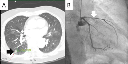
    Figure 1:  (A) 1.3cm-sized part solid nodule (black arrow) in the right lower
lobe at CT and (B) near total occlusion of the proximal LAD (white arrow) on
coronary angiography.

    Figure 2:  Capture of robot assisted LITA harvesting. Robot Debakey forceps
(white arrow), robot electrocautery spatula (black arrow), LITA (gray arrow).
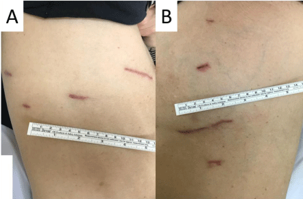
    Figure 3:  This patient’s wounds for 4th postoperative months. (A) Right side
for VATS and (B) left side for robot-assisted MIDCAB.
Discussion
Several surgical options can be considered to treat patients who have and coronary artery disease in the LAD and lung cancer in the right lower lobe: (1) percutaneous coronary intervention (PCI) and subsequent lobectomy, (2) simultaneous CABG and lobectomy through median sternotomy, (3) and complete minimally invasive surgery utilizing MIDCAB and VATS lobectomy. Staged PCI and lobectomy has limitations; if the PCI is performed before lobectomy, maintaining dual antiplatelet therapy (DAPT) increases risk of bleeding after lobectomy and discontinuing DAPT at perioperative period increases the possibility of acute stent thrombosis [1]. When performing simultaneous lobectomy and CABG via median sternotomy, lobectomy and mediastinal lymph node dissection may be problematic due to poor operative view, especially in case of lower lobectomy [2]. Moreover, invasiveness of median sternotomy can increase risk of postoperative complication particularly in the elderly. A previous study reported 13 cases in whom simultaneous lung resection for cancer and OPCAB were performed via median sternotomy; 3 of them suffered from major complications including postoperative bleeding, prolonged mechanical ventilation over 24 hours and prolonged air leakage [3]. Another study showed 6.7% of mortality (2 out of 30 patients) after simultaneous cardiac surgery and pulmonary resection via sternotomy [4]. Therefore, a minimally invasive approach such as simultaneous MIDCAB and VATS right lower lobectomy could be an effective treatment option in patients with LAD disease and lung cancer in the right lower lobe. In addition, robot-assisted LITA harvesting has advantage over direct vision LITA harvesting because over-retraction of rib and longer incision for direct vision could be avoided [5].
In conclusion, when surgical treatment is needed for LAD disease and lung cancer, simultaneous robot-assisted MIDCAB and VATS lobectomy might be a valuable option, particularly for elderly or frail patients.
References
- Levin GN, Bates ER, Bittl JA. ACC/AHA Guideline focused update on duration of dual antiplatelet therapy in patients with coronary artery disease, Circulation. 2016; 134: e123-e155.
- Morishita K, Kawaharada N, Watanabe T. Simultaneous cardiac operations with pulmonary resection for lung carcinoma. Jpn J Thorac Cardiovasc Surg. 2001; 49: 685-689.
- Dyszkiewicz W, Jemielity MM, Piwkowski CT, Perek B, Kasprzyk M. Simultaneous lung resection for cancer and myocardial revascularization without cardiopulmonary bypass (off-pump coronary artery bypass grafting). Ann Thorac Surg. 2004; 77: 1023-1027.
- Rao V, Todd TR, Weisel RD. Results of combined pulmonary resection and cardiac operation. Ann Thorac Surg. 1996; 62: 342-346.
- Ishikawa N, Watanabe G, Tomita S, Yamaguchi S, Nishida Y, Iino K. Robotassisted minimally invasive direct coronary artery bypass grafting. Thora CAB. Circ J. 2014; 78: 399-402.