Case Report
Austin J Surg. 2018; 5(9): 1154.
Spontaneous Splenic Rupture 48 Hours after Laparoscopic Sleeve Gastrectomy
Manuela T*, Anna F, Gabriele DA, Giuseppe S, Giovanni P, Riccardo R and Maria MG
Deartment of Bariatric Surgery, Humanitas Research Hospital, Italy
*Corresponding author: Trotta Manuela, Bariatric Surgery Unit, Humanitas Research Hospital - Via Alessandro Manzoni 56, Italy
Received: October 02, 2018; Accepted: November 06, 2018; Published: November 13, 2018
Abstract
Laparoscopic Sleeve Gastrectomy (SG) is the most commonly performed bariatric procedure in the management of morbid obesity. Post-operative splenic injury is a rare complication, and commonly seen as a delayed condition after surgery.
We report the first documented case of spontaneous splenic rupture two days after laparoscopic sleeve gastrectomy.
Keywords: Bariatric surgery; Laparoscopic sleeve gastrectomy; Splenic rupture; Postoperative complication
Case Presentation
A 55-year-old woman affected by dyslipidemia, glucose intolerance, severe gonarthrosis and mild gastro esophageal reflux, underwent laparoscopic Sleeve Gastrectomy (SG) at a BMI of 38 kg/ m2. The immediate postoperative course was uneventful, but severe left upper quadrant abdominal pain that radiated to left shoulder appeared on Postoperative Day (POD) [1].
Blood tests were normal. On physical examination, her pulse rate was 70 bpm, blood pressure was 130/70 mmHg, and oxygen saturation was 98% with ambient air.
Her abdomen was slightly distended, with mild diffuse tenderness and no peritoneal signs.
The persistent abdominal pain, lead us to perform an abdominal Computed Tomography (CT) scan. In the suspicion of gastric leak, also oral contrast was administered.
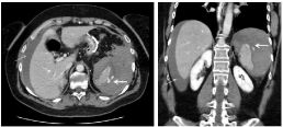
CT scan revealed free abdominal fluid and a perisplenic collection of 12 cm which was suspicious of splenic rupture. At the CT angiography, no active bleeding in the arterial phase was detected; instead, in the portal phase an intravenous contrast extravasation into peritoneal cavity was present (Figure 1).

    Figure 1:  CT scan, portal phase showing intravenous contrast extravasation
into peritoneal cavity (thick arrow), and hemoperitoneum (thin arrow).

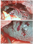
    Figure 2:  Intra-operative findings: subcapsular hematoma of the upper pole
of the spleen (thick arrows); multiple capsular tears (thin arrows).
No splenic nor portal venous thrombosis were found. No extraluminal air nor Gastrografin® was detected.
After CT scan, the patient gradually became hypotensive and tachycardic; she was rapidly resuscitated with an intravenous crystalloid bolus infusion and finally operated with explorative laparoscopy. An enlarged and fractured splenic upper pole with multiple capsular lacerations (Figure 2) and 2 L of hemoperitoneum were found. Indeed conversion to laparotomy and splenectomy was performed. The patient received 2 U of red blood cells and her postoperative recovery was uneventful (no intensive care unit required). Discharge from the hospital was on POD.
Pathologic study didn’t showed parenchymal abnormalities.
Discussion
Splenic injury is a well known intra-operative complication during bariatric surgical procedures; however it is also a possible but rare post-operative condition [2].
To our knowledge, there are few cases of splenic thrombosis, abscesses and capsular hematomas that lead to delayed splenic rupture, but no documented cases of early and spontaneous splenic fractures.
Salgado et colleagues [1] described the first case of post-operative splenic rupture as a complication of portal vein thrombosis eleven days after a SG, whereas Humenansky et al. [3] presented a fractured subcapsular hematoma secondary to air auto-dissection from pneumoperitoneum.
Also partial splenic ischemia may be involved as a mechanism of formation of delayed splenic abscesses that can rarely cause a rupture [4].
In the case we presented, splenic fracture was precocious and the etiology was still unclear.
No tears in the splenic capsule nor difficulties during short gastric vessels dissection were noted intra-operatively, however, at the emergency re-operation, sub capsular hematoma of the superior pole with capsular multiple fractures were found.
This, suggested us the possibility of an acute impaired venous drainage of the splenic upper pole causing an hypertensive hematoma, but we didn’t found venous thrombosis nor vascular abnormalities at CT scan.
Retrospective review of our collected data from 2010, showed this one as the first case of fractured spleen in 4130 consecutive laparoscopic SG.
More research and studies are still needed to determine the mechanism of this complication.
References
- Salgado N, Becerra P, Briceño E, Sharp A, Raddatz A. Splenic rupture as complication of sleeve gastrectomy. Surg Obes Relat Dis. 2012; 8: e72-e74.
- Lalor P, Tucker O, Szomstein S, Rosenthal R. Complications after laparoscopic sleeve gastrectomy. Surg Obes Relat Dis. 2008; 1: 33-38.
- Humenansky KM, Kopicky L, Opoku-Agyeman JL, Martinez CG. Splenic hematoma as a consequence of pneumoperitoneum. J Surg Case Rep. 2017; 10: 1-3.
- Nassour F, Schoucair NM, Tranchart H, Maitre S, Dagher I. Delayed Intra Splenic Abscess: a Specific Complication Following Laparoscopic Sleeve Gastrectomy. Obes Surg. 2018; 28: 589-593.