
Clinical Image
Austin J Surg. 2019; 6(9): 1182.
Gunshot Injury of the Atlanto-Axial Complex: An Unusual Solution to an Uncommon Contusion
Ohana N¹, Benharroch D²* and Sheinis D²
¹Orthopaedics, Meir Medical Centre, Kfar-Saba and Faculty of Medicine, Tel-Aviv University, Israel
²Pathology, Soroka University Medical Centre and Faculty of Health Sciences, Ben-Gurion University of the Negev, Israel
*Corresponding author: Daniel Benharroch, Pathology, Soroka University Medical Centre, 1, Rager Blvd, P.O.Box 151, Beer-Sheva 84101, Israel
Received: April 18, 2019; Accepted: April 25, 2019; Published: May 02, 2019
Clinical Image
This 29 y old male was admitted to our institution, following a gunfire assault.
On admission, the patient was alert and the vital signs were preserved. He had an entry wound in the anterior aspect of his neck. No exit wound was observed. The patient was neurologically intact.
Following an initial evaluation, the patient was transferred to radiology and had a computerized tomography of his head and neck, which showed the pictures enclosed.
The gun bullet was located next to the odontoid process of axis.
Although the patient was neurologically intact, concern of a potential dislodgement of the affected foreign body was considered. Potential plans of action included:
1. Leaving the bullet in place.
2. Fusing C1-C2 in order to prevent any movement of the bullet.
3. Removing the bullet.
Due to the patient’s young age, we decided to go for plan 3.
An anterior approach through the patient’s mouth was carried out. The musculature layers of the posterior oropharynx were divided longitudinally at the midline, exposing the anterior arch of C1. The bone was broken at its left aspect during the entry course of the bullet. We irrigated and removed the bone fragments. The breach was further extended with a bone drill to expose the bullet, as it lied adjacent to the odontoid process of axis. At this stage, we simply removed the bullet (Figures 1-10).
The bone defect was reconstructed with a bone graft, which was removed from the patient’s chin. The anterior arch of C1 was reconstructed with a 1.5 mm mini-plate (Synthes LTD) and the Figure 1: Imaging of the skull and cervical spine. A bullet is shown upon C1. wound was closed.
Following surgery, the patient was kept ventilated for 48 hours. Following the exhumation, he was allowed to step out of bed. He was later discharged.
Upon follow-up, the patient has full range of motion, no swelling is noted and he is neurologically intact.
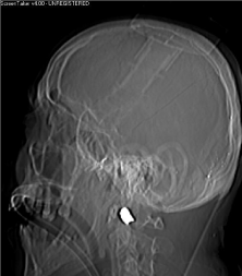
Figure 1: Imaging of the skull and cervical spine. A bullet is shown upon C1.
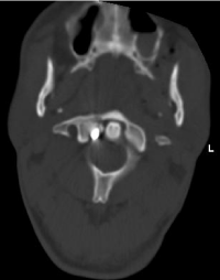
Figure 2: The bullet lies adjacent to the odontoid process of the axis.
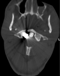
Figure 3: Fracture of left aspect of C1 with the bullet.
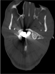
Figure 4: Fracture of left aspect of C1 with the bullet.
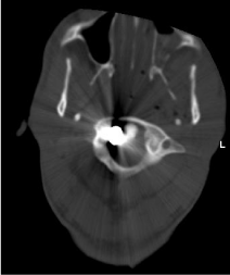
Figure 5: Fracture of left aspect of C1 with the bullet.
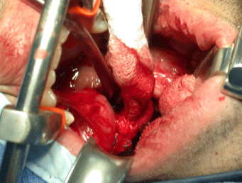
Figure 6: Anterior approach procedure through the mouth.
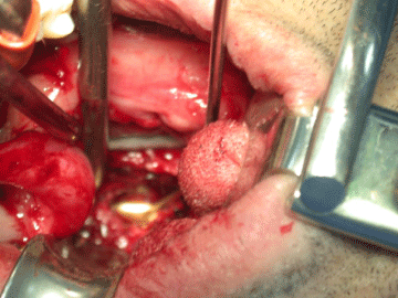
    Figure 7:  Following midline longitudinal muscle division, the bullet becomes
accessible.
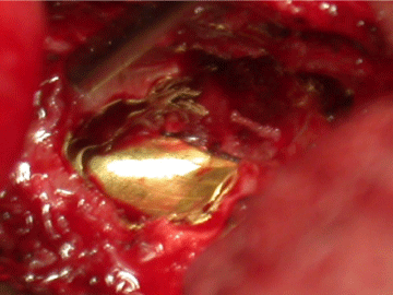
Figure 8: The bullet, uncovered.
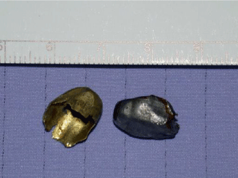
Figure 9: The bullet fragments.
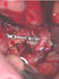
Figure 10: A prosthesis (Synthes LTD) replaces the anterior arch of C1.