Special Article - Aortic Surgery
Austin J Surg. 2019; 6(11): 1187.
Right Axillary Artery Cannulation and Selective Antegrade Cerebral Perfusion for Surgery of the Ascending Aorta: Initial Experience in a Low-Volume Center
Jacobzon E*, Kholod I, Tager S, Fink D, Merin O and Silberman S
Department of Cardiothoracic Surgery, Shaare Zedek Medical Center, Hebrew University of Jerusalem Medical School, Israel
*Corresponding author: Jacobzon E, Department of Cardiothoracic Surgery, Shaare Zedek Medical Center, Hebrew University Medical School, Jerusalem, Israel
Received: April 05, 2019; Accepted: May 06, 2019; Published: May 13, 2019
Abstract
Background: Arterial access for cardiopulmonary bypass in proximal aorta surgery is usually performed via the femoral, subclavian, or innominate arteries. We present our initial experience with axillary cannulation performed via the deltopectoral groove, with selective antegrade cerebral perfusion.
Methods: Included are 10 consecutive patients who underwent replacement of the ascending aorta with or without hemi arch: Type A dissection in 4, aortic aneurysm in 6. Right axillary artery is exposed with a deltopectoral groove incision. After division of the pectoralis minor and avoidance of the brachial plexus, an 8-mm graft is sewn end-to-side to the axillary artery and cannulated with a 22F Arterial cannula. Cardiopulmonary bypass is attained via right axillary artery and right atrium. After systemic cooling, the innominate artery is clamped and selective antegrade cerebral perfusion initiated at a flow of 10cc/kg/minute, with perfusion pressure around 60 mm/hg. Once the distal anastomosis of the ascending aorta graft is complete, the arterial clamp is removed from the innominate artery and systemic perfusion is renewed.
Results: Average systemic ischemic time was 36 minutes (range 24-60). Mean bypass time was 176 minutes (range 98-335). All patients made a noneventful recovery, with no incidence of strokes.
Conclusion: Arterial access using the right axillary artery via the right deltopectoral groove is simple and convenient. This artery is seldom involved in the disease process, and is reliably accessible, thus overcoming some of the major pitfalls of other venues. We have adopted this route as our preferred method for arterial cannulation for ascending aortic surgery.
Keywords: Axillary artery cannulation; Ascending aortic surgery; Cardiopulmonary bypass; Cerebral perfusion
Abreviations
CPB: Cardiopulmonary Bypass; DHCA: Deep Hypothermic Circulatory Arrest; MHCA: Moderate Hypothermic Circulatory Arrest; SACP: Selective Antegrade Cerebral Perfusion
Introduction
Surgery of the ascending aorta and aortic arch requires connection to Cardiopulmonary Bypass (CPB). In routine open-heart surgery, arterial access is most often attained via the distal ascending aorta. In cases in which the ascending aorta or arch are involved in the disease process, and particularly in cases of aortic dissection, there are a number of alternate sites for arterial access: the femoral artery, the innominate artery, the subclavian artery, and the axillary artery. When choosing between these options, we need to take into account the major causes of neurologic complications after proximal aortic surgery, which are embolic strokes and temporary neurologic dysfunction, usually due to global ischemia [1].
The right axillary artery is the distal continuation of the subclavian artery. It is easily accessible via an incision in the deltopectoral groove, far from any skeletal structure. It is rarely involved in the dissection process, which makes it an ideal cannulation site for surgery of the aorta. This approach also facilitates arterial access in complex cases such as re-operations. This technique has been previously described by Halkos et al. [2].
Axillary artery cannulation with Selective Antegrade Cerebral Perfusion (SACP) has become the standard of care for operations of the ascending aorta and the aortic arch in many leading centers worldwide. It enables continuous antegrade flow during cardiopulmonary bypass and uninterrupted SACP during systemic circulatory arrest. We have adopted this technique and during the past year have employed it in ten consecutive cases with excellent results. In this manuscript, we describe the technique and present our initial experience.
Materials and Methods
Included are 10 consecutive patients who underwent replacement of the ascending aorta with or without hemi arch replacement. Aortic pathology included: Type A dissection in 4, aortic aneurysm in 6.
Surgical technique:
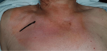
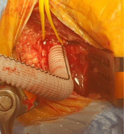
The right axillary artery is exposed via a deltopectoral groove incision (Figure 1). The pectoralis minor muscle is divided taking care to avoid the brachial plexus, and the axillary artery is exposed. After administration of heparin, the artery is clamped with a side-biting clamp and an 8-mm graft is sewn end-to-side to the axillary artery and then cannulated with a 22F arterial cannula (Figures 2a, 2b). Cardiopulmonary bypass is attained via right axillary artery and right atrium. After systemic cooling, the innominate artery is clamped proximally to enable selective antegrade cerebral perfusion initiated at a flow of 10cc/kg/minute, maintaining a perfusion pressure around 60 mm/hg [2,3]. Cerebral oxygen saturation is used to monitor brain perfusion. Replacement of the ascending aorta is performed in the usual fashion, and once the distal anastomosis is complete, the arterial clamp is removed from the innominate artery and systemic perfusion is renewed.

Figure 1: Deltopectoral groove incision site.

Figure 2a: Axillary artery is clamped (arrow) and an 8mm graft is sewn.

Figure 2b: Axillary artery is unclamped and the graft is filled.
Results
Total mean cardiopulmonary bypass time was 176 minutes (range 98-335). Average systemic ischemic time was 36 minutes (range 24-60). No patient died. All made a non-eventful recovery, with no incidence of stroke or any other neurologic deficit. Average utilization of packed red blood cells was 4.4 units per patient (range 2-10). Mean length of stay was 11 days (range 7-26). There were no local complications at the axillary cannulation site.
Discussion
In cases in which the ascending aorta or arch are involved in the disease process, and particularly in cases of aortic dissection, there are a number of issues that must be addressed. (i) The ascending aorta cannot reliably serve as the arterial cannulation site. (ii) Performance of the distal aortic anastomosis is best done with open technique under circulatory arrest. The latter requires special attention to brain protection, usually achieved by either accessing the right carotid artery in order to administer antegrade cerebral flow or administration of retrograde cerebral perfusion via the superior vena cava. Therefore, there are several common alternative options, such as the femoral, subclavian or innominate arteries.
The femoral artery is the most common alternative for arterial cannulation. It is easily accessible through a cut-down in the groin, with an option for venous cannulation as well. Nevertheless, it has several disadvantages. (i) It too can be involved in the dissection process, and entering its true lumen is not always obvious. (ii) Femoral cannulation provides retrograde arterial flow, which can promote neurological embolic events as well as reentries into a false lumen, especially when operating on dissected, atherosclerotic and diseased arteries. Thus, there is a degree of uncertainty as to the adequacy of organ perfusion. (iii) It cannot serve directly for cerebral perfusion, and using it for aortic surgery usually requires profound hypothermia and a separate cerebral perfusion method, either retrograde or direct antegrade.
The innominate artery can be accessed through the standard median sternotomy, usually with the mediastinal dissection extended above the innominate vein. By clamping it proximally, just at its bifurcation from the aortic arch, it can serve as a route for antegrade cerebral perfusion via the right common carotid artery. Its main disadvantage is that due to its proximity to the aortic arch it may be involved in the dissection process, and that ease of cannulation depends on the arch anatomy, which is not always favorable, especially with an enlarged and dissected ascending aorta.
The subclavian artery can also provide antegrade systemic flow and antegrade cerebral perfusion and is infrequently involved with the dissection process. Its anatomic location, partly beneath the clavicle, makes it less predictable and sometimes harder to access.
The majority of strokes after proximal aortic surgery are embolic in nature [3,4] and therefore are less likely to be affected by method of cerebral protection. Since the axillary artery is less often atherosclerotic, the risk of embolic stroke associated with manipulation of the artery is potentially reduced. The antegrade direction of flow is another factor that potentially reduces the risk of stroke.
Selective antegrade cerebral perfusion allows continuous brain perfusion thus prolonging the safe time of circulatory arrest. It also allows moderate rather than Deep Hypothermic Circulatory Arrest (DHCA) with favorable results, especially in elective cases [2,5].
Halkos et al presented a series of 271 patients comparing different venues for arterial cannulation for ascending aortic surgery. They employed DHCA with cooling to 18oC with either femoral or direct aortic cannulation in 66 patients, and SACP via the right axillary artery with moderate hypothermia (23oC) in 205. In their series, mortality was lower in the SACP group- 9% vs 23% [2]. Leshnower et al. published a series of 733 patients who underwent total arch replacement using Moderate Hypothermic Circulatory Arrest (MHCA) and unilateral SACP via the axillary artery. Core temperature at the onset of MHCA was 25.8oC with mean duration of circulatory arrest of 55 minutes. Overall incidence of stroke and transient neurologic deficit was 2.8% and 5.6%, respectively. Higher temperature was not found to be a significant risk factor for adverse events [5].
Conclusion
The right axillary artery provides a safe arterial access for cardiopulmonary bypass for first time as well as repeat cardiac surgery. It is easily adoptable even in centers with a low volume of aortic surgery. Inherent advantages include: (i) avoidance of aortic manipulation, (ii) antegrade bypass flow, and (iii) is a simple, safe and effective route for maintaining antegrade cerebral perfusion with moderate rather than deep hypothermia. As a result, we can achieve shorter cardiopulmonary bypass time and less postoperative complications. We have adopted this route as our preferred method for arterial cannulation for ascending aortic surgery. It can reduce major morbidity, especially in centers with low volume of aortic surgery.
References
- Hagl C, Ergin MA, Galla JD, Lansman SL, McCullough JN, Spielvogel D, et al. Neurologic outcome after ascending aorta-aortic arch operations: effect of brain protection technique in high-risk patients. J Thorac Cardiovasc Surg. 2001; 121: 1107-1121.
- Halkos ME, Kerendi F, Myung R, Kilgo P, Puskas JD, Chen EP. Selective antegrade cerebral perfusion via right axillary artery cannulation reduces morbidity and mortality after proximal aortic surgery. The Journal of Thoracic and Cardiovascular Surgery. 2009; 138: 1081-1089.
- Kazui T, Yamashita K, Washiyama N, Terada H, Bashar AHM, Suzuki Tohkura K, et al. Usefulness of antegrade selective cerebral perfusion during aortic arch operations. Ann Thorac Surg. 2002; 74: S1806-1809.
- Di Eusanio M, Schepens MAAM, Morshuis WJ, Di Bartolomeo R, Pierangeli A, Dossche KM. Antegrade selective cerebral perfusion during operations on the thoracic aorta: factors influencing survival and neurologic outcome in 413 patients. J Thorac Cardiovasc Surg. 2002; 124: 1080-1086.
- Leshnower BG, Kilgo PD, Chen EP. Total arch replacement using moderate hypothermic circulatory arrest and unilateral selective antegrade cerebral perfusion. J Thorac Cardiovasc Surgery. 2014; 147: 1488-1492.