Research Article
Austin J Surg. 2019; 6(12): 1190.
Genital Infections with Gardnerella vaginalis at Fann University Hospital (Dakar) and Saint-Louis (Senegal)
Diagne R¹*, Lo S², Dia ML³, Ndour J³, Ka R¹, Ngom B³ Cobar G³, Sarr H³, Niang AA³ and Sow AI³
¹UFR des Sciences de la Santé, Université de Thies BP 967 Thies, Sénégal
²UFR des Sciences de la Santé Université Gaston Berger BP234 Saint Louis, Sénégal
³Faculté de Médecine, Pharmacie, et Odontologie Université Cheikh Anta Diop BP 22254, Sénégal
*Corresponding author: Rokhaya Diagne, UFR des Sciences de la Santé, Université de Thies BP 967 Thies, Sénégal
Received: April 11, 2019; Accepted: May 08, 2019; Published: May 15, 2019
Abstract
Background: Bacterial vaginosis is a very common infection in women, especially during sexual activity.
We took stock of Gardnerella vaginalis infections in women received for vaginal swabs at the laboratories of two hospitals in Senegal: the National University Hospital Center (CHNU) in Fann and the Regional Hospital Center (CHR) in Saint Louis.
Material and Methods: Our study is retro-prospective over two years between January 1, 2013 and December 31, 2014. We worked on a total of 5928 vaginal samples.
We performed the measurement of vaginal pH, the potash test, the macroscopic and microscopic examinations on vaginal secretions.
Results: We obtained 1240 (21%) cases of Gardnerella vaginalis vaginosis, of which 826 (66.6%) originated from CHNU de Fann and 414 (33.4%) cases from the CHR of Saint-Louis.
The majority age group was [24-29 years old] with 317 cases.
The prevalence at Fann was 23% and 17% at the CHR of Saint-Louis.
More than 75% of the patients had whitish leucorrhea.
Conclusion: Given this prevalence, early diagnosis and effective treatment are essential to avoid clinical symptoms and complications related to this disease.
Keywords: Gardnerella vaginalis; Bacterial vaginosis; Genital infections
Introduction
Bacterial vaginosis is a common infection in women. In black Africa, it affects 20 to 50% of women [1-9]. It is responsible for 16 to 29% of cases of prematurity, chorioamniotitis, spontaneous abortions and low birth weights [3,4].
It results from a disorder of vaginal flora and an abnormal proliferation of commensal bacteria of the vagina (Gardnerella vaginalis, mycoplasms and anaerobic species). The precise origin of that disorder is unknown [10-16].
Gardnerella vaginalis is theincriminated bacterium in that condition. In some conditions, this bacterium can become pathogenic and lead to disorders of the vaginal flora. Its presence is higher in frequency (83 to 98% and in a much larger quantity than in the normal flora [8].
The aim of this study was to assess the prevalence of infections due to Gardnerella vaginalis in women received for vaginal swabs in the laboratories of two hospitals in Senegal.
Our study intends to elucidate the vaginal exsudat characteristics, but also to set up criteria of fast diagnosis. This will allow to better understand and ameliorate the diagnosis, but also show the place occupied by this condition in vaginal infections.
Matérials and Methods
This was a two-year retro-prospective study carried out between January 1 and December 31 of 2014 in women for which a vaginal swab was done at the Fann University Hospital and St Louis Regional Hospital.
Studied population
The studied population was made of patients admitted in the laboratory of these two above structures for patients who had a vaginal swab during that period of study.
The swab
The sample is taken on the vaginal walls or at the level of the posterior cul de sac. We made a PH vaginal measurement and a potash test, but also carried out micoscopic examinations and an identification of the germs involved in this infection.
Study of the samples
A microscopic examination gave us the colour, aspect and smell of the secretions. The microscopic examination, after a Gram coloration, highlighted the replacement of lactobacillus by a mixed flora: small coryneform bacilli to variable Gram suggesting Gardnerella bacilli to incurved variable Gram suggesting Mobiluncus.
A direct examination, after a Gram coloration, enabled to set up the type of flora.
    ARITAL    STATUS
    NUMBERS


    Single
    118


    Divorced
    29


    Engaged
    7


    Married
    617


    Widows

    14


    TOTAL
    785
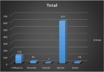
Table 1: Distribution of women according to marital status.
    GRAVIDITES
    NUMBERSS
    PERCENTAGES


    G0
    273
    35,4


    G1
    170
    22


    G2
    116
    15


    G3
    82
    10,6


    G4
    52
    6,8


    G5
    29
    3,8


    G6
    23
    3


    G7
    11
    1,4


    G8
    06
    0,7


    G9
    04
    0,5


    G10    and above
    02
    0,3


    Total
    772
    100
Table 2: Distribution of patients according to gravidity at Fann University Hospital.
    Colour of    leukorrhea
    FANN    University Hospital
    ST-LOUIS    Regional Hospital


    Whitish
    611
    316


    Greyish
    1
    3


    Haematic
    24
    14


    yellowish
    132
    70


    Milky
    4
    1


    Brown
    22
    7


    greenish
    21
    1


    Blackish
    1
    0


    Total
    816
    412
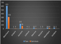
Table 3: Distribution of patients according to the colour of leukorrhea.
    Consistency    of leukorrhea
    Fann    University Hospital
    Saint-Louis    Regional Hospital


    Adherent
    47
    0


    Curd
    46
    23


    Thick
    292
    11


    Fluid
    91
    11


    Glairy
    121
    12


    Homogenous
    189
    352


    Soapy
    15
    2


    Normal
    9
    2


    Purulent
    3
    0


    TOTAL
    813
    413
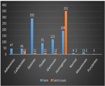
Table 4: Distribution of patients according to the consistency of leukorrhea.

    Figure 1:  Distribution of patients according to marital status at Fann
University Hospital.

Figure 2: Distribution of patients according to the colour of leukorrhea.

Figure 3: Distribution of patients according to the consistency of leukorrhea.
Results
Global results
We collected a total of 5928 samples of which 3493 (59%) came from the Fann University Hospital and 2435 (41%) from the St Louis Regional Hospital. We had 1240 cases of vaginosis to Gardnerella vaginalis of which 826 (66.6%) came from the Fann University Hosptal and 414 (33.4%) cases from the St Louis Regional Hospital.
The majority age group was [24 – 29 years old] with 317 cases.
The total prevalence of vaginosis to Gardnerella vaginalis was 21%. It was 23% at Fann University Hospital and 17% at St Louis Regional Hospital.
Sociodémographic results
Age distribution : The average age of patients was 31 years old.
At Fann University Hospital, portering was more common with age range [24-29 years old] with 28.1% followed by age ranges [30-35] years old, [18-23] years old, [36-41] years old, with respectively 25.3%, 17.5%, and 15%. At the St Louis Regional Hospital, the majority age group was [30-35] years old.
1. Distribution according to parity at Fann University Hospital
According to parity, the vaginosis to Gardnerella vaginalis was predominant in patients with less parity: 52% of women wit P0 parity; 17.7% for women with P1 parity; 13.2% for women with P2 parity; 7.4% for women with P3 parity; 3.8% for women with P4 parity; 3% for the P5; 1.2% for the P6 and less than 1% for each parity from P7 tp P14.
2. Other results
Flora IV types were the majority (73.3% in Fann) and (88.4% in St Louis).
The association rate Gardnerella vaginalis and Mobiluncus was 11.8% in Fann and 13.5% in St Louis.
Some association cases Gardnerella vaginalis and Chlamydia trachomatis were jotted down (2.7% in Fann and 17.8% in St Louis). However, it is worth noting that the Chlamydiae research was not systematic among all patients.
Discussion
During our study, the global prevalence of vaginosis to Gardnerella vaginalis in both structures was 21%. Our results are comparable to those obtained by Fofana (21.7%) in Bobo Dioulasso, but are inferior to those from Koueke in Cameroon (42%) [12].
Western writers jotted down rather badly matched prevalences between 15 and 60% [14,15].
The presence of Gardnerella vaginalis was higher in the age range between 24 and 35 years old in both structures. These results match with those of Faye-Kette who found out the same age ranges in Abidjan [5].
That situation could be explained by the fact that this age range is more active sexually. During that period, flora becomes favourable to infections [10].
Among women carrying Gardnerella vaginalis at vaginal smear, married women represented 78.5% of the cases against 15% for single women. These results match with those of Anagounou who found 60% of married women against 40% for single ones. On the contrary, Faye-Kette and coll. Found that single women were more numerous than married ones (59% against 41%) [2,5].
Leukorrhea was whitish in 76% and 75% of patients in St Louis and Fann respectively.The abundant leukorrhea was found in 46.2% in St Louis Regional Hospital and 45% in Fann University Hospital. while Faye-Keyette and coll. Found in greyish-white leukorrhea in the same proportions(76.5%) but abundance was more marked (62%) [6]. The distribution of bacterial vaginosis following the type of flora enabled to highlight the disorder of vaginal flora going with it. At Fann University Hospital, 11.8% of patients had the Gardnerella vaginalis and Mobiluncus spp association. In St Louis Regional Hospital, the association rate was 13.5%. These percentages are very low compared to those observed by Holst who had found bacerial vaginosis in more than 96% of women, and the presence of one or two Mobiluncus species [11].
The search for microplasm was carried out on 699 patients and that of Chlamydia on 629 ones.
In St Louis Regional Hospital, 36% of patients were positive to Gardnerella and Mycoplasma hominis, and as for Fann University Hospital, the association rate was 28.2%.
The association rate Gardnerella vaginalis and Ureaplasma urealyticum was of 62% at Fann University Hosptal and 36% at St Louis Regional Hospital. The M. hominis and U. urealyticum prevalences that we found in this study were higher to those reported by the literature, FAYE-KETTE while coll obtained 22% and 20% of women carrying Ureaplasma urealyticum of Mycoplasma hominis respectively [7]. The association rate Gardnerella vaginalis and Chlamydia trachomatis was of 17.8% at St Louis Regional Hospital and 2.7% at Fann University Hospital. This shows the difficulties in the treatment of this condition from multiple etiologies. As a result, a treatment targetting a single germ remains insufficient.
Conclusion
Gardnerella vaginalis constitutes the main agent of bacterial vaginosis. The diagnosis of this condition is easy to put in practice in laboratories. It can be responsible for complications like neonatal infections, repeated abortions, choroamnionitis and other obstetric infections. This study allowed us to show the place that occupy bacterial vaginosis in the infections of the uri-genital sphere.
With a 21% prevalence, we can conclude that vaginosis to Gardnerella vaginalis constitutes the first etiology of genital infections before candidiasis, trichomoniasis and infections to chlamydia and mycoplasms.
We recommend to strengthen the diagnosis to the laboratory of this condition so as to allow clinicians to have a good patient management but also to avoid complications related to that infection.
References
- Anagounou SY, Ndjoumessi G, Makoutode M. et al. Vaginose bactérienne chez la femme enceinte à Cotonou (Bénin). Méd Afr Noire. 1994; 41: 239-242.
- Askienazy-Elbhar M. Le diagnostic bactériologique des vaginoses bactériennes en pratique de ville. Rev Fr Gynécol Obstét. 1993; 88: 203-206.
- Bohot JM. Vaginose bactérienne. Paris, 2007 Extrait des mises à jour en Gynécologie Médicale volume 2007 publié le 12. 12. 2007.
- « www.cngof.asso.fr/d_livres/2007_GM_141_bohbot.pdf »
- Bresson L, Massoni S, Jailloux-Beaurain C, et al. Diagnostic de vaginose bactérienne par auto prélèvement vaginal pendant la grossesse: étude pilote. Rev Franc, Lab. 2006; 386: 45-48.
- Faye-Kette, Achi YH, Dosso M, Sylla –Koko DF, Akoua-Koffi. Test à la potasse et vaginite non spécifique à Abidjan. Soc Med Côte d’ivoire séance Avril. 1989.
- Faye-Kette A.V.H, Sylla-Koko OF, Cisse ALF et al. Aspects épidémiologiques et cliniques de la vaginose bactérienne à Abidjan. Méd Afr Noire. 1992; 39: 607 - 609.
- Faye Kette H, La Ruche G, Ali Napo L, Messou N, Viho I, Welfens-Ekra C, Dosso M et Sellati P. Genital mycoplasmas among pregnant women in Côte d’Ivoire, West Africa: prevalence and risk factors. Int J STD AIDS. 2000; 11: 599-602.
- Hill GB, St Claire KK, Gutman LT. Anaerobes predominate among the vaginal. Micro flora of prepubertal girls. Clin Infect Dis. 1995; 20: 269-270.
- Hillier SL, Lau RJ.Vaginal micro flora in post-menopausal women who have not received estrogen replacement therapy. Clin Infect Dis. 1997; 27: 123-126.
- Hillier SL, Martuis J, Krohn MA, Kiviat N, Holmes KK, Eschenbach DA. A case-control study of chorioamniotic infection and histology.Chorioamnionitis in prematurity. N Engl J med. 1988; 319: 972-978.
- Holst E. Reservoir of four-organism associate with bacterial vaginosis suggests Jack of sexual transmission. J Clin microbiology. 1990; 28: 2035-2039.
- Koueke P. La Gardnerellose bactérienne chez l'homme et chez la femme: traitement par l’association amoxicilline - métronidazole (Ospamox® - Supplin®) – Étude Préliminaire. Méd Afr Noire. 1996; 43: 384.
- Mitchell H. Vaginal discharge-cause, diagnosis, and treatment. BMJ. 2001; 328: 1306-1308.
- Morris M, Nicoll A, Simms L, Wilson J, Catchpole M. Bacterial vaginosis: a public health review. Br J Obstet Gynecol. 2001; 108: 439-450.
- Schmid GP Markowitz L, Joesoef. The epidemiology of bacterial vaginosis. Int J Gynaecol Obstet. 1999; 67: 17-20.