
Special Article – Surgery Case Reports
Austin J Surg. 2019; 6(12): 1192.
One-Stage Surgery for Two Conditions at the Same Vertebral Level: A Report of Two Cases and Literature Review
Gao X, Luo W, Sun Y, Chen Q and Gu R*
Department of Orthopedics, China-Japan Union Hospital of Jilin University, P.R.China
*Corresponding author: Rui Gu, Department of Orthopedics, China-Japan Union Hospital of Jilin University, No. 126 Xiantaida Street, Changchun 130033, P.R.China
Received: April 18, 2019; Accepted: May 17, 2019; Published: May 24, 2019
Abstract
Background: Cases of isthmic spondylolisthesis, degenerative lumbar kyphosis, and schwannomas have been widely reported, but cases involving more than one such condition occurring concurrently at a single vertebral level have rarely been reported.
Case Description: We present a case involving a female patient with isthmic spondylolisthesis combined with schwannoma that occurred at the same vertebral level, and a case involving a male patient with degenerative lumbar kyphosis combined with schwannoma that occurred at the same vertebral level. Preoperative examination, detailed physical examination, and general surgeryrelated consultation were performed before surgical treatment. The two patients underwent successful surgery, and a short-term follow-up showed that the symptoms were relieved.
Conclusion: Schwannoma coexisting at the same vertebral level with lumbar spondylolisthesis or kyphosis may be over looked; the necessary auxiliary examinations and detailed specialist examinations may help identify such lesions. After a comprehensive assessment of patient status, one-stage surgery for lumbar spondylolisthesis/kyphosis with schwannoma is feasible.
Keywords: Lumbar degenerative disease; Schwannoma; One-stage surgery; Case report
Abbreviation
T1WI: T1-weighted; T2WI: T2-weighted; MRI: Magnetic Resonance Imaging
Introduction
Isthmic spondylolisthesis is a clinically common degenerative disease of the spine, affecting 5% of adults [1]. Degenerative lumbar kyphosis is mainly caused by a decrease in the lordosis angle of the lumbar spine, potentially leading to lumbar kyphosis deformity. The average age of onset is often between 63.0 and 70.4 years, and the condition is related to poor lifestyle habits and heavy physical labor [2,3]. Schwannomas are common intradural extramedullary tumors originating from Schwann cells [4,5]. Approximately 95%–98% of schwannomas are benign. Ozawa et al. [6] reported that the incidence of schwannoma is 0.90 per 100,000 people per year, occurring mainly in the cervical and thoracic segments and rarely in the lumbosacral segment.
Isthmic spondylolisthesis, degenerative lumbar kyphosis, and schwannomas are common conditions. However, according to our knowledge, at the same vertebral level, there are no reports of simultaneous isthmic spondylolisthesis combined with schwannoma or degenerative lumbar kyphosis combined with schwannoma, which means there is a lack of treatment standards. We reviewed the available literature about patients with scoliosis and intraspinal anomalies. The classic surgical strategy was divided into two phases of surgery. The first stage treated the spinal canal lesions, and after 3 to 6 months, the second stage addressed spinal orthopedic issues [7,8]. Reports of one-stage surgery for scoliosis and intraspinal lesions are rare [9]. We report a case of isthmic spondylolisthesis combined with schwannoma which occurred at the same vertebral level, and a case of degenerative lumbar kyphosis combined with schwannoma which occurred at the same vertebral level.
Case Description
Ethical considerations
The ethics committee of China-Japan Union Hospital of Jilin University ruled that no formal ethics approval was required in this case. Written informed consent was obtained from all participants.
Case 1
The patient was a 56-year-old female farmer. She was admitted to our department with complaints of low back pain that she had been experiencing for 6 years and radiating pain in the left lower extremity that she had been experiencing for 3 years. Six years ago, there was no obvious cause of lumbar pain onset, which was aggravated with physical exertion and relieved by rest. No pain or numbness in the lower extremities were present at the time. Over the past 3 years, the low back pain gradually worsened and was accompanied by radiating pain and numbness in the left lower limb. The pain worsened with stretching, sitting, and standing positions and was relieved by lying down. The patient was physically healthy and denied any history of hypertension, diabetes, cancer, or surgery. Physical examination revealed a step between the spinous processes of L4-5; L4-5 and L5-S1 gaps were tender on palpation; there was increased pain with spinal extension, accompanied by radiating pain in the left lower extremity from the posterolateral side of the thigh to the lateral calf; decreased sensation over the left lateral calf and rear foot; grade 4 left hallux extensor muscle power; normal bilateral patellar and Achilles tendon reflexes; and a bilateral negative Babinski sign. Lumbar X-ray showed L5-S1 isthmic spondylolisthesis (grade II). Computed tomography (CT) showed a L5-S1 left intervertebral foramen stenosis with isthmus fracture, and a space-occupying lesion was seen behind the L5 vertebral body. The space-occupying lesion showed normal or decreased signal intensity on T1-weighted (T1WI) magnetic resonance imaging (MRI), increased intensity on T2-weighted (T2WI) sequences, and uneven enhancement on contrast-enhanced MRI (Figure 1). The patient was diagnosed with isthmic spondylolisthesis and schwannoma in the general practice consultation. After a comprehensive evaluation of the patient’s condition, L4-5 posterolateral fusion, L5-S1 inter body fusion, and subdural tumor removal were performed during one-stage surgery under mixed intravenous and inhaled anesthesia. The tumor was completely removed during surgery (Figure 2). The patient’s symptoms were significantly relieved after surgery. Postoperative pathological findings revealed the lesion was a schwannoma (Figure 3). After 1 year of follow-up, the L5-S1 fusion was in good condition, and the neurological function returned to normal (Figure 4).
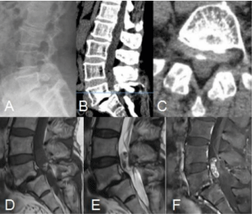
    Figure 1: Preoperative imaging.
A: Lumbar X-ray shows L5-S1 grade II spondylolisthesis. B and C: CT shows
left L5-S1 intervertebral foramen stenosis and L5 isthmus fracture. D and
E: MRI shows normal or decreased signal intensity in the T1WI sequence
and increased signal intensity in the T2WI sequence. F: Enhanced scanning
shows uneven enhancement.
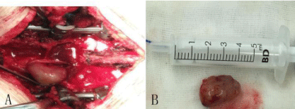
    Figure 2: Intraoperative images.
A: Tumor located in the intradural area. B: The tumor has a complete capsule,
which is an elliptical solid occupying place.
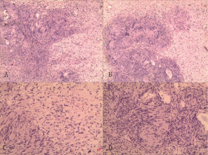
    Figure 3: Hematoxylin and eosin-stained sections of formalin-fixed and
paraffin-embedded intradural xtramedullary schwannoma specimen.
A and B: Low-magnification (×40) H & E section showing typical Antoni A hyper
cellular areas composed of spindle cells arranged in fascicular and sheetlike
patterns, with nuclear palisading (circles). C and D: High-magnification
(×100) H & E sections showing elongated, spindle-shaped tumor cells with
mild nuclear atypia. There are no necrotic areas.
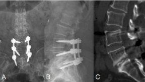
    Figure 4: Postoperative images.
A and B: The position of the internal fixation is good. C: Satisfactory L5-S1
intervertebral fusion.
Case 2
A 64-year-old male farmer was admitted to the hospital with pain in the lumbar and left hip areas for 5 years, which worsened over the last 3 days. Five years ago, there was no obvious cause of lumbar pain onset; the pain was relieved by bed rest. There was no pain or numbness in his lower limbs. Over the past 3 days, the pain in the lower back suddenly increased, accompanied by radiating pain in the left hip, which was relieved when lying down, and worsened when changing position. He denied any history of hypertension, diabetes, cancer, or surgery. Examination revealed slight bulging and tenderness over L2-3. The left hallux extensor muscle power was grade 4, and the muscle power of all major muscle groups of the lower limbs was grade 5. The left patellar tendon reflex was brisk, the Achilles reflexes were diminished, and the Babinski sign was negative bilaterally. Lumbar X-ray radiography showed slight lumbar scoliosis, and lateral radiography showed kyphosis of L2-3. CT showed a spontaneous fusion tendency of the L2-3 vertebral body and loss of height of the L2-3 disc. The space-occupying lesions showed normal or decreased intensity signals in T1WI MRI and increased intensity signal both on the T2WI sequences and in the fat-suppressed images (Figure 5). The patient underwent one-stage open reduction internal fixation and intraspinal tumor removal under mixed intravenous and inhaled anesthesia. The tumor was completely removed during surgery (Figure 6). The patient’s symptoms were significantly relieved after surgery. Postoperative pathological findings suggested that the lesions were schwannomas (Figure 7). After 6 months of follow-up, the fused segments were well aligned, symptoms were significantly improved, and nerve function was normal (Figure 8).
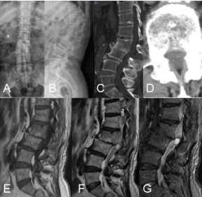
    Figure 5: Preoperative images.
A: Lumbar vertebrae X-ray shows mild lumbar scoliosis, B: Lumbar lateral
X-ray shows kyphosis between L2-3. C and D: CT shows a spontaneous
fusion tendency of the L2-3 vertebral bodies. E and F: MRI shows normal or
reduced signal intensity in the T1WI sequence and increased signal intensity
in the T2WI sequence. G: Fat-suppressed image shows increased signal.
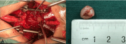
    Figure 6: Intraoperative images.
A: Tumor located in the intradural area. B: The tumor showing a complete
capsule, which is an elliptical solid occupying place.
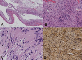
    Figure 7: Hematoxylin and eosin- stained sections of formalin-fixed and
paraffin-embedded intradural extra medullary schwannoma specimen.
A: Low magnification (×40) H&E staining shows typical Antoni A cell area and
cystic changes. B: Medium magnification (×100) H&E staining shows that
the tumor cells were arranged in a fence. C: High magnification (×400) H&E
staining shows a nucleus with mild abnormalities and interstitial edema. D:
Immunohistochemistry (×200) shows positive S-100 staining.
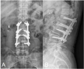
    Figure 8: Postoperative Images.
A and B: Good internal fixation.
Discussion
Isthmic spondylolisthesis, degenerative lumbar kyphosis, and schwannoma are common diseases, but at the same vertebral level, isthmus spondylolisthesis combined with schwannoma and degenerative lumbar kyphosis combined with schwannoma have rarely been reported. The two patients underwent detailed examination and evaluation and completed one-stage surgery under general anesthesia with good postsurgical results.
Isthmic spondylolisthesis and schwannoma can cause lumbar pain and abnormal nerve function in the lower extremities. In addition to low back pain, the patients presented with decreased sensation over the left lateral lower leg and rearfoot, and the muscle power of the extensor hallucis longus was grade 4. These findings were consistent with L5-S1 intervertebral foramen stenosis. Therefore, we believe that the intervertebral foramen stenosis caused by isthmic spondylolisthesis (grade II) and L5 nerve root compression was the main cause of symptoms in these patients. In cases of degenerative lumbar kyphosis and intraspinal schwannomas in patients with lumbar pain and postural changes after left hip pain, we believe that the lumbar pain arises mainly from lumbar instability, but hip pain may be caused by nerve sheath tumor or nerve compression at the lumbar spine.
Treatment of spinal deformities and canal abnormalities requires a surgical approach. Two-stage surgery leads to scar formation, affecting normal anatomy and causing difficulties in the second surgery, thereby increasing the likelihood of complications and possibly delaying wound healing [10]. Moreover, performing a second-stage surgery increases the risk of iatrogenic nerve damage [10,11]. Mehta et al. [11] compared patients undergoing one-stage surgery with patients undergoing two-stage surgery, and the results showed that the risk of complications in the two-staged surgery group was significantly higher.
Once an abnormality in the spinal canal is discovered at the level of spinal deformity, the following questions must be considered: (1) Does the abnormality require treatment? (2) Can surgical treatment improve spinal deformity? (3) If surgery is needed, what is the appropriate time to perform surgery? Our one-stage intraspinal space surgical treatment was performed due to excessive tumor volume, since a simple decompression and fusion may lead to neurological deficits; additionally, in two-stage surgery, first-stage tumor resection will lead to local scar formation and, as a result, the second-stage fusion process will be more difficult. Before surgery, we evaluated imaging data and predicted the complications that could occur during intraoperative correction. We considered that the spinal deformity of the two patients would be significantly improved postoperatively. For the timing of surgery, we considered that the earlier the surgery, the better the prognosis would be. One-stage surgery is safer, more effective, requires a shorter hospital stay, improves spinal orthopedics, and presents no significant surgical complications [11].
However, this study has some limitations. First, we have only two cases of spinal deformity and intraspinal schwannoma. We need more patients to assess the safety and efficacy of one-stage surgery. Second, because the surgery is relatively new, we still need to monitor for new neurological symptoms and orthopedic deficits. Third, one of the patients did not have an enhanced MRI before surgery. Although it did not lead to missed diagnosis or misdiagnosis, the relevant examination should be completed before surgery to avoid a similar situation.
Conclusion
Preoperative examination is of great significance to establish an accurate diagnosis and avoid a missed diagnosis. One-stage surgery for the treatment of isthmic spondylolisthesis combined with schwannoma and kyphosis combined with schwannoma is effective and feasible.
Funding
This study was funded by the Department of Finance of Jilin Province (grant number 3D517N903430 to Rui Gu).
References
- Murray MR, Skovrlj B, Qureshi SA. Surgical treatment of isthmic spondylolisthesis. Clin Spine Surg. 2016; 29: 1-5.
- Jang JS, Lee SH, Min JH, Han KM. Lumbar degenerative kyphosis: radiologic analysis and classifications. Spine (Phila Pa 1976). 2007; 32: 2694-2699.
- Bae JS, Jang JS, Lee SH, Kim JU. Radiological analysis of lumbar degenerative kyphosis in relation to pelvic incidence. Spine J. 2012; 12: 1045-1051.
- Ando K, Imagama S, Ito Z, Kobayashi K, Yagi H, Hida T, et al. How do spinal schwannomas progress? The natural progression of spinal schwannomas on MRI. J Neurosurg Spine. 2016; 24: 155-159.
- Viereck MJ, Ghobrial GM, Beygi S, Harrop JS. Improved patient quality of life following intradural extra medullary spinal tumor resection. J Neurosurg Spine. 2016; 25: 640-645.
- Ozawa H, Aizawa T, Kanno H, Sano H, Itoi E. Epidemiology of surgically treated primary spinal cord tumors in Miyagi, Japan. Neuroepidemiology. 2013; 41: 156-160.
- Jankowski PP, Bastrom T, Ciacci JD, Yaszay B, Levy ML, Newton PO. Intraspinal pathology associated with pediatric scoliosis: A ten-year review analyzing the effect of neurosurgery on scoliosis curve progression. Spine (Phila Pa 1976). 2016; 41: 1600-1605.
- Winter RB, Haven JJ, Moe JH, Lagaard SM. Diastematomyelia and congenital spine deformities. J Bone Joint Surg Am. 1974; 56: 27-39.
- Samdani AF, Asghar J, Pahys J, D’Andrea L, Betz RR. Concurrent spinal cord untethering and scoliosis correction: case report. Spine (Phila Pa 1976). 2007; 32: 832-836.
- Hamzaoglu A, Ozturk C, Tezer M, Aydogan M, Sarier M, Talu U. Simultaneous surgical treatment in congenital scoliosis and/or kyphosis associated with intraspinal abnormalities. Spine (Phila Pa 1976). 2007; 32: 2880-2884.
- Mehta VA, Gottfried ON, McGirt MJ, Gokaslan ZL, Ahn ES, Jallo GI. Safety and efficacy of concurrent pediatric spinal cord untethering and deformity correction. J Spinal Disord Tech. 2011; 24: 401.