Special Article - Oral & Maxillofacial Surgery
Austin J Surg. 2019; 6(13): 1194.
Surgical Anatomy of Mandibular Third Molar
Gupta S1*, Khan TA1, Attarde H3 and Narula J4
1Department of Oral & Maxillofacial Surgery, Jaipur Dental College, India
2YMT Dental College and Hospital, Navi Mumbai, India
3Student of International Dental Program, Moldova State University of Medicine and Pharmacies, Moldova
*Corresponding author: Gupta S, Department of Oral & Maxillofacial Surgery, Jaipur Dental College, Dhand, Jaipur, Rajasthan 302028, India
Received: April 29, 2019; Accepted: May 22, 2019; Published: May 29, 2019
Abstract
A proficient knowledge of oral anatomy is mandatory to facilitate an uncomplicated removal of lower third molars. This article addresses basic anatomic structures that are in close relationship to lower third molars. Pertinent bones, muscles, blood supply, nerve innervations, which may be encountered during Trans alveolar extraction of third molar are reviewed; along with their surgical implications. Caution must be exercised while performing surgery in this area and thus making the reader more enlightened on this aspect. This article was constructed by reviewing English literature from 1993 to till date. This review article concludes that a more comprehensive study should be conducted in this purview of mandibular third molar.
Keywords: Surgical anatomy; Third molar; Nerve injury; Complication of third molar extractions
Introduction
Surgical removal of impacted third molar is one of the common surgical procedures carried out in the oral and maxillofacial surgery set up. Surgical management of impacted third molar is difficult because of its anatomical position, poor accessibility, and potential injuries to the surrounding vital structures, nerves, vessels, soft tissues, and adjacent teeth during surgeries [1].
To overcome these hurdles of surrounding anatomical structures of third molar; we embarked on this review of literature to make the reader more aware of the surgical anatomy as well as possible complications associated with these structures. For a comprehensive review, we have categorised this anatomy into hard tissues and Soft tissues.
Hard Tissues
Bones
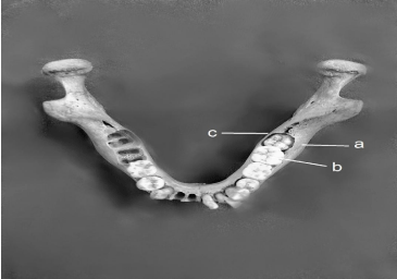
When the mandible is viewed anteriorly, it is seen that the ramus tends to flare out buccal from the distal aspect of the third molar region. Mandibular third molar is situated at the distal end of the body of the mandible at the junction of mandibular body and the ramus. This junction constitutes a line of weakness and The lower third is embedded between a thick buccal cortical plate which is buttressed by the external oblique ridge and on the lingual side is present the comparatively narrower lingual cortical plate where is connection with relatively thin ramus [2] (Figure 1).

    Figure 1: Key: (a) External oblique ridge, (b) Adjacent second molar, (c) Thin
lingual plate.
Surgical implications: Fracture may occur if excessive force is applied at the junction mandibular body and the ramus in elevating an impacted mandibular third molar [2].
The external oblique ridge on the buccal side is a bulky prominence and actually impedes and resists buccal traction of mandibular third molar [2].
Adjacent teeth
The teeth adjacent to lower third molar are mandibular second molar, which are present mesial to them. They can be healthy, heavily restored, carious or missing.
Surgical implications: Clinical and radiographic evaluation of the patient is essential to avoid damage to proximal teeth when removing third molars. Second mandibular molar teeth with large restorations, crowns, or caries may be damaged during the removal of third molars [3]. Teeth with large restorations or carious lesions are always at risk of fracture or damage upon elevation. Correct use of surgical elevators and bone removal can help prevent this occurrence. The incidence of damage to adjacent restorations of the second molar has been reported to be 0.3% to 0.4% [4].
Retromolar foramen
The RMF is an inconstant foramen situated in the central portion of the retromolar fossa, which is bounded by the anterior border of ramus of the mandible and temporal crest. The foramen receives a canal of variable depth that normally arises from the mandibular canal behind the lower third molar, which is regarded as the retromolar canal (RMC). Normal morphological findings of the human mandible and its possible variations that occur have attracted special interest in the recent years in the field of odontos tomato logical surgeries. One such anatomical variation, which draws special attention in clinical dental practice, is the Retro molar foramen in the retromolar trigone (RMT). The trigone is bounded medially by temporal crest, laterally by the anterior border of the ramus and anteriorly by the base of third molar tooth. The Retro molar foramen has generally been neglected in anatomical text books and this has been rarely studied or reviewed [5].
in the dental literature. The frequency of Retro molar foramen has been reported in different populations showing a wide varying incidence between 3.2% and 72% the distance between the 3rd molar and Retro molar foramen is being within the short range of 4–11 mm [5].
Surgical implications: This close relation of Retro molar foramen with mandibular third molar could lead to damage of the structures traversing through RMF during the third molar extraction and could be a reason for postoperative hematomas due to rupturing of the vessels in Retro molar foramen [5].
Soft Tissues
Buccinator muscle
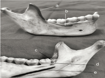
The buccinator muscle is a plain, square-shaped bilateral mimic muscle, which composes the mobile and adaptable portion of the cheek. It is frequently referred to as an accessory muscle of mastication because of its role in chewing food and swallowing and compressing the cheeks against the molars, as well as its use for whistling, sucking, and blowing [6] (Figure 2).

    Figure 2: Key: (a) Temporalis muscle, (b) Buccinator muscle, (c) Temporalis
tendon, (d) Superior pharyngeal constrictor muscle( SPCM), (e) Mylohyoid
muscle.
Although the buccinators muscles pierces the distal portion of the retro molar pad, the precise origin of the attachment is the periosteal layer and the underlying alveolar bone is completely bare of muscle attachments [7].
Surgical implications: Extensive stripping of mucoperiosteum beyond the muscle attachment at the vestibule results in troublesome swelling, hematoma and postoperative discomfort [7].
Mylohyoid muscle
Mylohyoid is a triangular, flat muscle that forms the floor of mouth. It is attached to the entire mylohyoid line and meets the other fellow in a midline fibrous raphe that extends from symphysis mention to hyoid bone. The inferior surface is covered by platysma, and anterior belly of digastric is closely related to it [8]. This broad sheet like muscle forms a sling inferior to the tongue, defining the boundary between the sublingual space and the submandibular space. The sublingual space is super medial to the mylohyoid muscle and lateral to the genioglossus and geniohyoid muscles in the oral cavity [9].
At the third molar area, the mylohyoid muscle is inferior to the lingual nerve. At the posterior attachment of the mylohyoid muscle to the mandible, the lingual nerve makes an anteromedial turn and travels superficial to the mylohyoid muscle. Mylohyoid serves functions like chewing, swallowing, respiration and phonation [8].
Surgical implications: As the lower third molar is encased predominantly by lingual alveolus, the roots lie in proximity to or their apices may even perforate the lingual plate. Attempting elevation of such roots, may lead their displacement through the thin lingual cortex and deflection backwards below the mylohyoid muscle and along the medial surface of mandible into the so-called lingual pouch. Difficulty may be experienced in retrieving of such dislocated roots [7].
Pogrel recommended that the operator place his or her thumb underneath the inferior border of the mandible in an attempt to direct the tooth back along the lingual surface of the mandible [10]. The lingual gingiva may be reflected as far as the premolar region and the mylohyoid muscle incised to gain access to the submandibular space and deliver the tooth. In this approach, care should be taken to avoid injury to the lingual nerve in this anatomic region.
Temporalis tendon
The temporalis muscle is a fan-shaped muscle that originates from the temporal fossa and inserts on to the coronoid process of the mandible. The tendon starts high within the muscle in the form of a broad tendinous lamina that gradually thickens and converges on the coronoid process. The temporalis tendon wraps around the medial and anterior surfaces of the coronoid, extending down onto the anterior mandibular ramus. A small portion is seen on the lateral surface of the coronoid. Most of the tendon is found on the medial aspect and extends inferiorly toward the buccinator line [11] (Figure 2).
Surgical implications: A buccal approach to third molar region interferes with the lowest part of the temporalis tendon, i.e., the part of tendon has to be sectioned to be able to remove the buccal and distal bone covering of the molar. Moreover, during retraction of buccal flaps, a downward and/or buccal traction with retractor may lacerate the periosteum in the flap or extend the base of flap beyond the external oblique ridge, leading to increase in pain, swelling, and trismus. When any muscle is damaged, a pain reflex is stimulated. This condition is called “muscle guarding,” which results when muscle fibers engender pain and when they are stretched. This pain causes the muscles to contract, resulting in loss or range of motion [12].
Superior pharyngeal constrictor muscles
Gray described the origin fibers of the superior pharyngeal constrictor muscle at mandible to be found at the lingual surface of the mandible. The site of attachment is at, or below, the convexity of the mandibular lingual crest above a point called the mylohyoid line and medial to the third molar tooth. The muscle fibers serve to anchor and stabilize the wall of the superior pharynx in respiration, phonation, and narrows the pharynx in deglutition [13] (Figure 2).
Surgical implications: Edwin [13] stated that there is a possibility of injury to superior pharyngeal constrictor muscle while performing transalveolar extraction of lower third molar.
Lingual Nerve (LN)
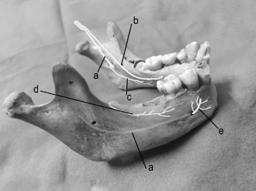
The lingual nerve is a branch of the mandibular nerve, provides somatosensory innervation of the lingual mucosa. Jointly with the chorda tympani nerve fibers, a branch of the facial nerve, it provides information to the anterior two thirds of the dorsum of the tongue and preganglionic parasympathetic innervation of submandibular and sublingual secretory glands [14]. Its location is medial and anterior to the inferior alveolar nerve. It continues under the gingival sulcus of the lingual mucosa superficially to the surface of the gland and submandibular ganglion. It ends as a sublingual nerve located immediately below the tongue mucosa. Lingual nerve is reported to be at the level of the alveolar crest or higher in 17.6% of cases. In the retromolar region, the distance from the lingual alveolar crest to the lingual nerve is averaged at 4.45 mm and ranges between 3.01 mm to 2.28 mm in the third molar area. Its proximity to the third molar lingual cortical plate, separated from the cortex only by the periosteum, and its variable anatomyare the most important risk factors to be considered by the oral surgeon during surgical removal of the third molar [15,16] (Figure 3).

    Figure 3: Key: (a) Inferior alveolar nerve (IAN), (b) Lingual nerve, (c)
Mylohyoid nerve (d) Buccal nerve, (e) Mental nerve.
Surgical implications: One of the important prevention strategies to avoid iatrogenic lingual nerve injury is obtaining a thorough knowledge of lingual nerve anatomy and topography. The inclination of the alveolar lingual plate in addition to the prominence of the alveolar process influence the lingual nerve position at the lower third molar region. Lingual nerve will be more cranial in position if there is a short distance between mandibular ascending ramus and the lower third molar. Therefore, marked mandibular atrophy could be an important risk predictor for lingual nerve injury during surgery in the area with any lingual flap manipulation. The lingual split technique in addition to lingual flap retraction is associated with an increased risk of temporary nerve damage compared with the buccal approach plus and the simple buccal approach. Although tooth sectioning does not increase the incidence of lingual nerve damage, it could be a possible risk factor for lingual nerve injury during extraction of lower third molar. The nerve may be directly cut by the rotary instrument during tooth sectioning. Therefore, sectioning two thirds of the lower third molar followed by using straight elevator to complete the separation would prevent the risk of lingual nerve injury. Furthermore, lingual nerve damage is also associated with removal of periradicular bone at the distolingual or lingual sites. Suture -Although there is little data reporting on damage to the lingual nerve due to suturing, it is evident that the lingual nerve could be damaged by direct trauma from the needle if it is inserted too apically. The nerve can also be compressed if included within the flap of the tissue adapted by the suturing. Over exuberant and excess tissue bites for suturing must be avoided and the use of compressive/occlusivemaneuvers for surgical haemostasis must take into account the position of the nerve. Interestingly if the nerve and associated blood supply has been damaged during surgery, the first sign is significant bleeding which is difficult to control. Methods to stop the bleeding (suturing, haemostat, cautery) can worsen the status of an already damaged nerve [15].
The frequency of lingual nerve injuries during third molar removal ranges from 0.2 to 22% presenting with temporary sensory deficits and 0-2% having permanent sensory disturbances [17].
A systematic review has shown no difference in permanent LN injury rates whether a lingual retractor was used or not. There is no relation between the incidence of LN injury and the angulations of the mandibular third molar and LN injury [15].
Inferior Alveolar Nerve (IAN)
This nerve arises as a branch of mandibular nerve (V3) in the infratemporal fossa. It appears at the inferior border of the inferior head of lateral pterygoid muscle, courses downward, and enters the mandibular foramen; it gives numerous sensory branches that innervate the mandibular bone. These small nerves are in association with small vessels in neuro vascular channels. The inferior dental nerve can run as once unit through the mandibular canal until it reaches the premolar region, where it divides into the mental and incisive nerves [18] (Figure 3).
Most third molar roots in close proximity to the IAN canal were buccal (45%), in line with the canal (39%), lingual (10%) and 6.4% were interradicular; 20% of roots were more than 6 mm from nerve, 3% 0–1 mm, 48% 0 mm with cortication, and 29% 0 mm with cortical break [19].
Surgical implications: The relationship of mandibular third molar to inferior alveolar nerve must be considered when surgical removal is contemplated surgical planning and proper informed consent depend on detailed knowledge of the positional relationships in this area the more intimate the relationship of the inferior alveolar neurovascular bundle to the roots of the teeth, the more likely nerve damage to occur. Patients must have an understanding of the potential consequences of IAN damage, and options such as leaving the tooth alone and coronectomy should be considered in cases in which the likelihood of IAN damage is significant [20].
Third molar surgery-related inferior alveolar nerve injury is reported to occur in up to 3.6% of cases permanently and 8% of cases temporarily. If the tooth is closely associated with the IAN canal radiographically (e.g. superimposed on the IAN canal, darkening of roots, loss of lamina dura of canal, deviation of canal). 20% of patients having these teeth removed are at risk of developing temporary IAN nerve injury and 1–4% is at risk of permanent injury [19].
Buccal Nerve
This nerve descends between the two parts of the lateral pterygoid muscle, medial to the ramus of the mandible, and then passes laterally across the external oblique ridge distal to the third molar, to supply the cheek. The sensory distribution is variable but includes the lower posterior buccal sulcus and gingivae, and an area of cheek mucosa. As the nerve crosses the external oblique ridge it is composed of between one and five branches, the lowest of which may be over 1 cm below the deepest concavity of the ridge [21].
Surgical Implications: Anatomical studies carried out on the long buccal nerve show that it is at risk during the initial incision for many third molar procedures. Branches of it are probably frequently cut during the incision process, but the effects are generally not noted but a few patients complain of complete anaesthesia of the cheek; the incidence of this complication has not been reported [21].
Mylohyoid Nerve
The Mylohyoid Nerve (MHN) is a branch of the Inferior Alveolar Nerve (IAN), which arises above the mandibular foramen. The nerve then passes downward and anteriorly within the mylohyoid groove on the medial surface of the mandible, gives branches that provide motor innervations to the mylohyoid and anterior belly of the digastric muscles [22] (Figure 3).
It has been analysed that the MHN might have a role in the sensory innervations of the chin [23].
Surgical implications: The mylohyoid nerve injured during lingual retraction of flap in third molar surgery. The study, published in 1992 by Carmichael, found injury to the mylohyoid nerve to be as high as 1.5%, probably due to lingual retraction [24]. There is very little evidence in the literature demonstrating injury to the mylohyoid nerves following the removal of impacted mandibular third molars. The nerves most commonly affected are the inferior alveolar and lingual nerves.
Facial artery
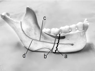
The facial artery normally arises from the external carotid artery, just above the lingual artery, at the level of greater cornu of hyoid bone in the carotid triangle. It then passes upwards and forwards medial to the ramus of the mandible. It passes deep to the superficial part of the submandibular salivary gland making a characteristic loop, winds around the base of the mandible to enter the face at anteroinferior angle of the masseter muscle. On the face, it runs upwards and forward, laterals to angle of the mouth, and terminates as angular artery [25] (Figure 4).

    Figure 4: Key (a) Facial artery, (b) Facial vein, (c) Inferior alveolar artery, (d)
Inferior alveolar vein.
Facial vein
The facial veins, coursing with or parallel to the facial arteries, are valveless veins that provide the primary superficial drainage of the face. Tributaries of the facial vein include the deep facial vein, which drains the pterygoid venous plexus of the infratemporal fossa. Inferior to the margin of the mandible, the facial vein is joined by the anterior (communicating) branch of the retromandibular vein. The facial vein drains directly or indirectly into the IJV. At the medial angle of the eye, the facial vein communicates with the superior ophthalmic vein, which drains into the cavernous sinus [26] (Figure 4).
Inferior alveolar artery
The inferior alveolar artery is a branch of the maxillary artery, one of the two terminal branches of the external carotid. Prior to entering the mandibular foramen, it gives off the mylohyoid artery. In approximately the first molar region, it divides into the mental and incisal branches. The mental branch exits the mental foramen and supplies the chin and lower lip, where it eventually will anastomose with the submental and inferior labial arteries [27].
Inferior alveolar Vein
The inferior alveolar vein forms from the merging of its dental branches, alveolar branches, and mental branches in the mandible, where they also drains into pterygoid plexus of veins. The dental branches of inferior alveolar vein drain the pulp of mandibular teeth by way of each tooth’s apical foramen. The alveolar branches of the inferior alveolar vein drain the periodontium and gingival of the mandibular teeth [28].
Life-threatening haemorrhage resulting from the surgical removal of third molars is rare. However, copious bleeding from soft tissue is relatively common. One source of bleeding during the surgical removal of third molars is the inferior alveolar artery or vein [29].
The facial artery and the anterior facial vein, cross the inferior border of the, mandible just anterior to the masseter muscle and have a close relationship to the second molar [7].
Haemorrhage from mandibular molars is more common than bleeding from maxillary molars (80% and 20%, respectively), because the floor of the mouth is highly vascular. The distal lingual aspect of mandibular third molars is especially vascular and an accessory artery in this area can be cut leading to profuse bleeding [29].
Surgical implications of vessels: Inferior alveolar artery and vein can be cut during sectioning of third molars, leading to profuse bleeding [29].
It is possible to cut facial artery and vessels if the scalpel should slip when making a buccal cut in the third molar region; therefore, it is advisable always to begin the incision in the depth of the sulcus and direct the blade upwards towards the teeth [7].
Buccal fat pad
It is a deep fat pad located on either side of the face and is surrounded by the following structures:
1. Anterior- Angle of the mouth
2. Posterior- Masseter muscle
3. Medial- Buccinator muscle
4. Lateral- Platysma muscle, subcutaneous tissue, and skin
5. Superior- Zygomaticus muscles
6. Inferior- Depressor angular or is muscle and the attachment of the deep fascia to the mandible.
Surgical implications: The buccal fat pad is a structure that may be encountered when removing impacted third molars. Deep incisions distal to the maxillary tuberosity may cause herniation of the buccal extension. The unexpected herniation of the buccal fat pad is alarming. However, this complication is typically innocuous [3,30].
Spaces Involved with Lower Third Molars
The accidental displacement of a lower third molar or one of its root fragments is not common during extraction, nevertheless a wellrecognized complication that is frequently mentioned in textbooks [31].
The tooth/root fragments may get displaced into the following spaces:
Sublingual space
The sublingual area is a triangular virtual space, located in the floor of the mouth, above the mylohyoid muscle, under the free portion of the tongue. The lateral limit of the sublingual space is the muscle complex hyoglossus-styloglossus, while the anterior limit is the genioglossus muscle. Important morphologic structures are observed in the sublingual space, such as the duct of the submandibular salivary gland, branches of the lingual artery, and the lingual and hypoglossal neural bundles [32].
Submandibular space
The submandibular spaces are considered to be the anterior extension of Para pharyngeal space [33]. It is bounded anteromedially; the floor formed by mylohyoid muscle, which is covered by loose areolar tissue and fat, Postero-medially, the floor formed by hyoglossus muscle, Supero-laterally, medial surface of mandible below the mylohyoid ridge, Antero-superiorly, anterior belly of digastricus, Postero-superiorly, posterior belly of digastric, stylohyoid and stylo-pharyngeous muscle, laterally by platysma and skin [33]. It contains superficial lobe of salivary gland and submandibular lymph nodes, facial artery and vein [33].
Pterygomandibular space
The inferior head of the lateral pterygoid muscle bind the pterygomandibular space superiorly, laterally by the medial aspect of the ramus of the mandible and anteriorly it is continuous with the recess formed by the lateral pterygoid and temporalis muscles. The interpterygoid fascia [34] binds it medially and posteriorly.
Moorthy have earlier suggested that an extra oral approach may provide better access if the displacement is deeper into the substance of the medial pterygoid muscle or inferiorly into the submandibular space. A combination of intraoral and extra oral approaches may be needed to retrieve tooth and root fragments in certain situations [35].
Surgical implication of spaces: It usually occurred when the tooth was located lingual, where there is fenestration of the lingual cortical plate with root exposure, and where surgical technique may be inadequate. Displaced fragments may vary in size and may appear in different tissue spaces [31].
Conclusion
This review article makes us understand the need for more studies regarding a good assessment of region around mandibular third molar to avoid any mishap and treat these trans alveolar extractions with great care.
References
- Vibha singh, Khonsao Alex, Pradhan R, Mohammad S, Singh N. Techniques in the removal of impacted mandibular third molar: A comparative study. European journal of general dentistry. 2013: 2: 25-30.
- Chakravarthy C. Textbook of oral and maxillofacial surgery. 2nd edition. Paras, New delhi, Hyderabad. 2011; 178.
- Wayland J. Impacted Third molars. 1st edition. Wiley Blackwell. 2018; 57.
- Chiapasco M, De cicco L, Marrone G. Side effects and complications associated with third molar surgery. Oral surg oral med oral pathol. 1993; 76: 412-420.
- Potu BK, Kumar V, Salem AH, Abu-Hijleh M. Anatomy Research International. 2014.
- Baghele ON, Baghele MO. Buccinator Muscle Repositioning: A Case Report. Compendium. 2012; 33: 1-6.
- Chakravarthy C. Textbook of oral and maxillofacial surgery. 2nd edition. Paras, New delhi, Hyderabad. 2011; 178.
- Jha S, Khorwal G. A Rare Case of Accessory Nerve To Mylohyoid Communicating With Lingual Nerve And Its Clinical Implications. International Journal of Anatomy and Research. 2018; 6: 5550-5553.
- Yamamoto MO, Nakajima K, Tsuji Y, Otonari T, Curtin HD, Okano T, et al. Imaging of the Mylohyoid Muscle: Separation of Submandibular and Sublingual Spaces. American Journal of Roentgenology. 2010; 194: 431-438.
- Bouloux GF, Steed MB, Vincent J. Perciaccante. Complications of Third Molar Surgery. Oral Maxillofacial Surg Clin N Am. 2007; 19: 117–128.
- Boahene KD, Farrag TY, Ishii L, Byrne PJ. Minimally Invasive Temporalis Tendon Transposition. Facial Plast Surg. 2011; 13: 8-13.
- Balakrishnan G, Narendar R, Kavin T, Venkataraman S, Gokulanathan S. Incidence of Trismus in Transalveolar Extraction of Lower Third Molar. J Pharm Bio allied Sci. 2017; 91: 222–227.
- Ernest EA, Ernest MW. George Salter. Superior Pharyngeal Constrictor Muscle Pain. Practical pain management. 201 6.
- Zur KB, Mu L, Sanders I. Distribution pattern of the human lingual nerve. Clin Anat. 2004; 17: 88-92.
- Alali Y, Mangat H, Marco F. Caminiti. Oral Health. 2018.
- Holzle FW, Wolff KD. Anatomic position of the lingual nerve in the mandibular third molar region with special consideration of an atrophied mandibular crest: an anatomical study. Int J Oral Maxillofac Surg. 2001; 30: 333-338.
- Kale TP, Pandit VS, Patil S, Pawar V, Shetty N. Lingual Guttering Technique for Removal of Impacted Mandibular Third Molars. J Int Oral Health. 2014; 6: 9-11.
- Carl E, Misch, Hamza A, Abbas. Contemporary Implant Dentistry.3rd edition. Mosby. 2008; 497.
- Renton T. Prevention of Iatrogenic Inferior Alveolar Nerve Injuries in Relation to Dental Procedures Dent Update. 2010; 37: 350-363.
- Pogrel MA. partial odontectomy. Oral And Maxilla Fac Clinic Of North Am. 2007; 19: 85-91.
- Loescher R, Smith KG, Robinson PP. Nerve Damage and Third Molar Removal. Dent Update. 2003; 30: 375-382.
- Clark S, Reader A, Beck M, Meyers WJ. Anesthetic efficacy of the mylohyoid nerve block and combination inferior alveolar nerve block mylohyoid nerve block. Oral Surg Oral Med Oral Pathol Oral Radiol Endo. 1999; 87: 557-563.
- Guyot L, Layoun W, Richard O, Cheynet F, Gola R. Alteration of chin sensibility due to damage of the cutaneous branch of the mylohyoid nerve during genioplasty. J Oral Maxillofac Surg. 2002; 60: 1371-1373
- Carmichael FA, McGowan DA. Incidence of nerve damage following third molar removal: A West of Scotland Surgery Research Group study. British J Oral Maxillofac Surg. 1992; 302: 78-82.
- Venugopal SV, Rao V, Ravindra Kumar B, Bhasin G. Relationship Between The Facial Artery and Sub Mandibular Salivary Gland. Int J Anat Res. 2014; 23: 597-600.
- Moore, Keith L, Dalley, Arthur F. Clinically Oriented Anatomy, 5th Edition. 951
- Randolph R. Resnik. Carl E Misch. Misch’s Avoiding Complications in Oral Implantology. Chapter-7, Elsevier. 2018; 267-293.
- Margaret J, Susan FB, Herring W. Illustrated anatomy of head and neck.5th edition. Elsevier. Chapter 6. 147
- Wayland J. Impacted Third molars. 1st edition. Wiley Blackwel. 2018; 4.
- Wayland J. Impacted Third molars. 1st edition. Wiley Blackwell. 2018; 7.
- Huang IY, Wu CW, Worthington P. The Displaced Lower Third Molar: A Literature Review and Suggestions for Management. J Oral Maxillofac Surg. 2007; 65: 1186-1190.
- Silveira RJ, Garcia RR, Botelho TL, Franco A, Silva RF. Accidental Displacement of Third Molar into the Sublingual Space: A Case Report. J Oral Maxillofac Res. 2014; 5.
- Pankaj S, Richa T, Sudeep G, Archana RR, Mudit A, Abhishek S. Journal of Science. 2015; 54: 235-237.
- Vora MM, Nagargoje P. Displacement of a mandibular Third molar in the Pterygomandibular space -a case report. Journal of Applied Dental and Medical Sciences. 2015; 13: 67-69.
- Olayinka AM, Olutayo J, Olakusehin LA, Olurotimi FS. Iatrogenic displacement of impacted mandibular third molar into the submandibular space complicated by submasseteric abscess. Afr J Trauma. 2016; 51: 19-22.