Special Article – Surgery Case Reports
Austin J Surg. 2019; 6(14): 1196.
Deceased-Donor Liver Transplantation for Budd Chiari Syndrome Long Segmental Thrombosis of the Inferior Vena Cava with Extensive Collateral Circulation
Liu ZX, Zhu JQ, Ma J, Kou JT, Li XL and He Q*
Department of Hepatobiliary and Pancreatic Splenic Surgery, Capital Medical University, China
*Corresponding author: He Q, Department of Hepatobiliary and Pancreatic Splenic Surgery, Capital Medical University, China
Received: May 02, 2019; Accepted: May 31, 2019; Published: June 07, 2019
Abstract
Introduction: Liver transplantation is a rescue treatment for patients with Budd-Chiari Syndrome (BCS) who develop into an end-stage liver disease. Very few of these patients have been reported to have long-segmental thrombosis of the Inferior Vena Cava (IVC). Herein, we presented a patient with total obstruction of the IVC blocked by a long-segmental thrombus.
Case Presentation: A 26-year-old BCS patient with complete obstruction of the IVC and the hepatic veins was transferred to our hospital. Deceased donor liver transplantation was scheduled for him. During the operation, the donor’s suprahepatic IVC was anastomosed to the recipient’s thoracic IVC, and the sub hepatic IVC was sutured as the iliac veins and the renal veins were drained into the superior vena cava smoothly through collateral circulation. Postoperative course was uneventful.
Conclusion: Before liver transplantation, the surgeons should assess the BCS patient’s vascular anatomy and hemodynamic cautiously, and the patients could benefit from reconstruction of the venous outflow.
Keywords: Budd-Chiari Syndrome; Liver transplantation; Long-sengmental thrombosis; Collateral circulation
Introduction
Budd-Chiari Syndrome (BCS) is an infrequent clinical disease resulting from obstruction of the hepatic venous outflow tract anywhere from the hepatic venules to the right atrium, including the Inferior Vena Cava (IVC) and the hepatic veins (HVs) [1]. Currently, according to the patient’s individual status, a step-wise treatment strategy has been proposed and widely adopted, which contains anticoagulation, thrombolysis, percutaneous recanalization, transjugular intrahepatic portosystemic shunt (TIPS) and surgical shunt [2]. It has been reported that up to 10-20% BCS patients still develop into liver function failure after the step-wise treatment [3]. Therefore, liver transplantation is the remaining rescue treatment in these patients. Notably, the five-year survival rate of BCS patients who underwent liver transplantation can reach as high as 80% [4,5]. Most of BCS patients progress to liver failure because of the partial or short-segmental obstructed IVC and/or HVs. On the other hand, very few patients have a complete obstruction of the IVC. Herein, we presented a patient with total obstruction of the IVC blocked by a long-segmental thrombus, where the iliac veins and the renal veins were drained into the superior vena cava smoothly through collateral circulation.
Case Report
Clinical features and laboratory findings
A 26-year-old Chinese male with an end-stage liver disease was transferred to our hospital. He complained of an abdominal distending pain for 2 months, accompanied with low fever and poor appetite. He denied any past medical history. Physical examination merely revealed massive as cites.
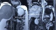
Laboratory findings were: hemoglobin level, 16.8g/dL; platelet count, 179,000/mm3; albumin level, 3.2g/dL; alanine aminotransferase (ALT), 514U/L; aspartate aminotransferase (AST), 500U/L; total bilirubin level, 105umol/L; direct bilirubin level, 62umol/L; international normalized ratio, 2.13; Prothrombin activity, 35.8%; normal levels of the tumor markers and renal function. The MELD score and the Child-Pugh score were 22 and 11, respectively. Abdominal enhanced magnetic resonance imaging (MRI) showed the IVC was utterly obstructed by a long-segmental thrombus from the suprahepatic IVC to the common iliac vein (Figure 1); both of the iliac veins and the renal vein were drained into the superior vena cava via the varicose azygos-hemi-azygos vein; besides, the right, middle and left hepatic veins were all completely thrombosed. As the patient developed liver failure, deceased donor liver transplantation (DDLT) was scheduled.

    Figure 1: The IVC was totally blocked by a long-segmental thrombus from
the suprahepatic IVC to the common iliac vein. IVC, inferior vena cava.
Surgery
The orthotropic liver transplantation was performed. During the operation, approximately 5 L of ascites and extensive collateral circulation were observed in the abdominal cavity. The thrombosed retrohepatic and suprahepatic IVC were resected together with the liver. Then, the donor’s suprahepatic IVC was anastomosed with the recipient’s thoracic IVC directly. The recipient’s distal subhepatic IVC was sutured because there was no blood backflow. The patient’s circulatory system was stable during the operation.
Postoperative condition
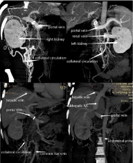
Postoperative course of the patient was uneventful. By the end of the first postoperative week, abdominal enhanced Computed Tomography (CT) was done, which demonstrated that all the anastomotic vessels including the suprahepatic IVC were patent (Figure 2). Abdominal Magnetic Resonance Angiography (MRA) was performed every 6 month to make sure the anastomotic vessels were patent. Oral aspirin was taken per day. The laboratory results were all within the normal range during the 2-year follow-up.

    Figure 2: Both of the iliac veins and the renal vein were drained into the
superior vena cava via the varicose azygos-hemi-azygos vein.
Discussion
BCS patients have to undergo liver transplantation due to cirrhosis and liver failure, which result from liver congestion. Congestion is induced by increased sinusoidal pressure, owing to poor clinical outcomes and/or technical failure of the step-wise treatment [6]. Most of BCS patients just have a short-segmental or partial obstruction of the IVC. Sun reported that more than 80% of BCS patients developed into partial and short-segmental obstruction of the IVC [7]. Besides, only a small number of BCS patients will develop IVC thrombosis. Menthe’s retrospective analysis showed that among BCS patients who received liver transplantation, less than 17% of them had IVC thrombosis [5]. Therefore, it is even rarer for BCS patients to have a long-segmental complete thrombosed IVC. According to Karaca’s study on BCS patients who underwent living donor liver transplantation (LDLT), only 3 patients progressed to a complete obstruction of the IVC from the suprahepatic IVC to the renal veins [8]. While, in our case, the IVC was totally blocked from the suprahepatic IVC to the common iliac vein. The venous blood flow from both kidneys and the lower extremities was drained into the superior vena cava via the varicose azygos-hemi-azygos venous system.
In addition, the most difficult challenge is reconstruction of venous outflow during liver transplantation. Various methods could be taken for reconstruction: venous drainage may be reconstructed on the already stenosed and/or thrombosed IVC [8]; surgeons could prune a wide de novo orifice on the IVC [9]; cadaveric vessels could be used as an alternative [9,10]; in selected patients, the graft’s hepatic vein could be anastomosed to the right atrium directly [11]. In this patient, as the native IVC was totally blocked by a long-segmental thrombus, we had to remove the thrombosed retrohrpatic and suprahepatic IVC simultaneously with the liver. Considering the normal renal function and normal lower limbs, we sutured the subhepatic IVC without changing the patient’s original hemodynamics of lower limbs and kidneys. Due to the long range of the thrombus, we anastomosed the graft’s suprahepatic IVC to the recipient’s thoracic IVC directly. Postoperative results of this case present an excellent surgical effect during the follow-up.
In conclusion, BCS patient’s collateral vascular situation and the extent of venous obstruction should be adequately evaluated before surgery, so the surgeons could determine how to reconstruct the blood vessels.
References
- Hidaka M, Eguchi S. Budd–Chiari syndrome: Focus on surgical treatment. Hepatol Res. 2017; 47: 142-148.
- Seijo S, Plessier A, Hoekstra J, Dell’Era A, Mandair D, Rifai K, et al. Good long-term outcome of Budd-Chiari syndrome with a step-wise management. Hepatology. 2013; 57: 1962-1968.
- Valla DC. Budd-Chiari syndrome/hepatic venous outflow tract obstruction. Hepatol Int. 2018; 12: 168-180.
- Segev DL, Nguyen GC, Locke JE, Simpkins CE, Montgomery RA, Maley WR, et al. Twenty years of liver transplantation for Budd-Chiari syndrome: a national registry analysis. Liver Transpl. 2007; 13: 1285-1294.
- Mentha G, Giostra E, Majno PE, Bechstein WO, Neuhaus P, O’Grady J, et al. Liver transplantation for Budd–Chiari syndrome: A European study on 248 patients from 51 centres. J Hepatolo. 2006; 44: 520-528.
- Simonetto DA, Yang HY, Yin M, Assuncao TM, Kwon JH, Hilscher M, et al. Chronic passive venous congestion drives hepatic fibrogenesis via sinusoidal thrombosis and mechanical forces. Hepatology. 2015; 61: 648-659
- Sun YL, FuY, Zhou L, Ma XX, Wang ZW, Wu Y. Staged management of Budd-Chiari syndrome caused by co-obstruction of the inferior vena cava and main hepatic veins. Hepatobiliary Pancreat Dis Int. 2013; 12: 278-285.
- Karaca C, Yilmaz C, Ferecov R, Iakobadze Z, Kilic K, Caglayan L, et al. Living-Donor Liver Transplantation for Budd-Chiari Syndrome: Case Series. Transplantation Proceedings. 2017; 49: 1841-1847.
- Ara C, Akbulut S, Ince V, Karakas S, Baskiran A, Yilmaz S. Living donor liver transplantation for Budd-Chiari syndrome: Overcoming a troublesome situation. Medicine. 2016; 95: 5136.
- Cetinkunar S, Ince V, Ozdemir F, Ersan Y, Yaylak F, Unal B, et al. Living- Donor Liver Transplantation for Budd-Chiari Syndrome-Resection and Reconstruction of the Suprahepatic Inferior Vena Cava With the Use of Cadaveric Aortic Allograft: Case Report. Transplantation Proceedings. 2015; 47: 1537-1539.
- Kazimi M, Karaca C, Ozsoy M, Ozdemir M, Apaydin AZ, Ulukaya S, et al. Live donor liver transplantation for Budd-Chiari syndrome: anastomosis of the right hepatic vein to the right atrium. Liver Transplantation. 2009; 15: 1374- 1377.