Case Report
Austin J Surg. 2019; 6(17): 1205.
Hydronephrosis in a Patient with Bladder Hydatid Disease - Case Report and Review of the Literature
Klacz J*, Krukowski J, Szczypior M, Czajkowski M and Matuszewski M
Department of Urology, Medical University of Gdansk, Poland
*Corresponding author: Klacz J, Department of Urology, Medical University of Gdansk, 17 Smoluchowskiego St, Gdansk, Poland
Received: July 18, 2019; Accepted: August 12, 2019; Published: August 12, 2019
Abstract
Hydatid disease is an endemic zoonosis which can affect humans causing serious health problems. We present a case report of 47-year-old patient who presented to the urology department with right kidney hydronephrosis due to bladder calcified cyst. Surgical treatment and pathological examination showed that the patient suffered from bladder hydatiosis.
Keywords: Hydatid disease; Hydroneprosis; Hydatid cyst; Urinary tract; Bladder cyst; Tapeworm
Introduction
Hydatid disease (HD) is an endemic zoonosis caused by the larval form of Echinococcus tapeworm: E. granulosus and E. multilocularis, which lives in the gut of canines and other carnivorous animals [1]. Uncommonly humans may become accidental intermediate hosts by ingesting tapeworm eggs, mostly by fruits and vegetables not washed carefully enough.
All organs in the human body may be affected by hydatid disease. Excluding common localizations such as lungs and liver all other organs are considered as uncommon. Urinary tract involvement is extremely rare and accounts to 1-5% of all cases [2].
Case Report
A 47-year-old man presented to the urology department with a right flank pain, urgency, nycturia and complaints of abdominal discomfort. On questioning, patient said that few years earlier he was treated because of hydatid disease. He also had undergone cholecystectomy and left-sided nephrectomy because of purulent inflamation and afunction of left kidney before. He denied any hematuria.
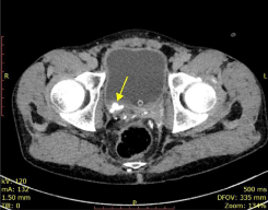
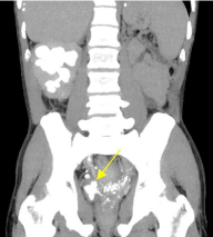
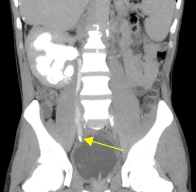
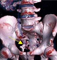
Physical examination revealed positive Goldflams’s sign over the right kidney. His renal function was normal. Abdominal ultrasonography showed right hydronephrosis and multiple calcified objects, especially in liver, right kidney and bladder wall. A contrastenhanced computed tomography (CT) scan of the abdomen and pelvis revealed multiple locations of calcified cysts. One of them was located in bladder wall (Figure 1 – arrow), pressing on distal part of the right ureter (Figure 2, 3, 4 – arrow). The patient has remained on anti-hydatid therapy for 18 years. The disease was estimated as stable and he was qualified for the operation. After difficult preparation of extensive fibrosis embracing ileum and pelvic walls the excision of the cyst with small part of distal ureter and bladder wall was done followed by ureterocystoneoanastomosis with double J stent placement. An 18Fr Foley catheter was retained for 12 days after the operation. Three days after the surgery patient was reoperated because of small intestine obstruction and perforation. The patient recovered and was discharged after 14 days of hospitalization. Double J stent was removed 6 weeks later.

Figure 1: Calcified object in back wall of the urinary bladder.

Figure 2: Narrowing of the ureter with calcified object around it.

Figure 3: Narrowing of the ureter with calcified object around it.

    Figure 4 – 3D: CT reconstruction showing large calcified cyst pressing on
distal part of the right ureter.
Pathological examination showed that the excised cyst was one of many of hydatid cysts. Antiparasitic treatment with albendazole was continued in outpatient clinic. Control ultrasonography and CT after 8 months showed no signs of recurrent hydronephrosis.
Discussion
Hydatid disease is caused by the larval form of E. granulosus and E.multilocularis[1]. Like other cestodes, echinococcal species have both intermediate and definite hosts. Usually canines (dogs, foxes) are the definitive hosts [3]. The small adult worm which lives in the canine bowel pass their eggs in the feces which are consumed by intermediate hosts – horses, sheep, humans. The embryos from the eggs penetrate the intestinal mucosa, enter the portal circulation and reach various organs [3,4]. The larvae develope into fluid filled hydatid cysts. The hydatid cysts commonly affect organs such as: liver (60%), lungs (20%), kidneys (4%), spleen (3%), brain (3%), soft tissue and bone (2%). Bladder hydatid cysts are extremely rare [2, 3]. It is endemic in many countries where dogs, sheeps and human live in close contact. However, as a result of emigration of infected persons, cases do occur in non-endemic areas, where diagnosis is often delayed because the true diagnosis is not considered [1].
In general a conclusive diagnosis of hydatiosis is often made intraoperatively. The diagnosis is usually late because of the slow evolution of the disease and the absence of symptoms and apparently good health of the patient. Eosinophilia is not always seen. Immunological assays are now being used for detection of specific antibodies, circulating antygen and immune complexes. However, there is no test that is higly sensitive and specific, particularly for cystic HD [1]. Imaging by CT or ultrasound is considered as the main tools for diagnosis. Serology and other tests are considered complementary [6]. The advantages of ultrasound are that it is simple, inexpensive, and provides instant results.
Early urinary tract hydatid disease (HD) can be difficult to diagnose due to the slow growth of cysts, the presence of non-specific and subtle clinical manifestations and the rarity of daughter vesicles in the urine, the defining sign of this disease. That is why careful history taking is crucial. Urologists are usually confronted with advanced stages when long time after infection cysts or peri-cystic fibrosis affect urinary tract. Lower back pain and kidney tenderness are the most common symptoms of urinary tract HD, but this symptoms are clearly not specific, so urinary tract HD is often overlooked in differential diagnoses. From a renal point of view, hydatidosis can cause haematuria or low back pain. Also, glomerulonephritis mediated by immune complexes and secondary amyloidosis have been described [5]. Few cases of retrovesical hydatid cyst have been published, but still HD in that area is a rare finding (0.1%-0,5% of cases) [5].
Although urinary tract HD has a relatively low incidence, misdiagnosis can have major health consequences [6,7]. Especially dangerous is the sponateous rupture of an abdominal or pelvic cyst or dissemination by the Douglas pouch during a surgical procedure. It usually gives nonspecific urinary symptoms. Less than 15% of retrovesical hydatid cysts can fistulise, mostly to the bladder, with hydatiduria being a pathognomonic sign of vesicular cystic fistula [5].
CT scanning has several advantages for the diagnosis of urinary tract HD in that it provides visualization of cyst wall calcification and intracystic infection, and also allows identification of low-density and high-density areas, which is important for the differentiantion of a HD cyst from a urinary tract abscess or tumor [6].
Surgery is the treatment of choice for hydatid disease [1,2,3,6,8]. The experience with preoperative and postoperative therapy with antiparasitic drugs showed that albendazole is recommended to reduce the risk of dissemination during surgery and to prevent recurrence [8]. Administration of this agent for one week to one month before surgery may reduce the intraoperative tension of the HD cyst, prevent HD spread during surgery, and may kill or reduce the activity of Echinoccocus larvae [6]. Continuous use of albendazole for 3 months after surgery may also reduce postoperative recurrence, especially when cystic fluid has spread during surgery [6].
Conclusion
Hydatiosis of urinary tract is uncommon and may present with various symptoms leading to diagnostic difficulty and management problems. Being aware of its possible presence is important especially in the endemic areas. Surgical excision of the cyst remains the treatment of choice with long pharmaceutical prophylaxis.
References
- Papanikolaou A. Osseous hydatid disease.Trans R Soc Trop Med Hyg. 2008; 102 (3): 233-8.
- Sallami. Intravesical hydatid cyst. Urology. 2005; 66: 1110.e7-1110e8.
- Kanagal DV, Hanumanalu LC. Hydatid cyst of urinary bladder associated with pregnancy: a case report. Arch Gynecol Obstet.2010; 282 (1): 29-32.
- White AC, Weller PF. Cestodes. Harrison’s principles of internal medicine.17th edn. Pp. 1339-1340.
- Sánchez-Rodríguez L, Serra-Díaz C, Domenech-Iglesias A, Sánchez- Sanchís M, Alvarez-Avellán L, López-Menchero R, et al. Primary retrovesical hydatidosis as a cause of chronic kidney disease.Nefrologia. 2013; 33(2): 285-6.
- Huang M, Zheng H. Clinical and demographic characteristics of patients with urinary tract hydatid disease. PLoS One. 2012; 7 (11): e47667.
- Ozbey I, Aksoy Y, Polat O, Atmaca AF, Demirel A. Clinical management of hydatid disease of the urinary tract. J Int Med Res. 2002; 30: 346-352.
- Feki. Multiply unusual locations of hydatid cysts including bladder, psoas muscle and liver. ParasitolInt. 2008; 57(1):83-6.