Special Article – Surgery Case Reports
Austin J Surg. 2019; 6(18): 1210.
A Case of Midface Reconstruction via a Nasolabial Perforator Flap, Gone through Extreme Venous Outflow Problem
Alp E1* and Leman Damla E2
1Istanbul Teaching and Research Hospital, Department of Plastic, Reconstructive and Aesthetics Surgery, Turkey
2Istanbul University Faculty of Medicine, Departmen of General Surgery, Turkey
*Corresponding author: Alp ERCAN, Istanbul Teaching and Research Hospital, Department of Plastic, Reconstructive and Aesthetics Surgery, Haseki Sultan Mah. Cevdetpasa Caddesi 94/12 Fatih/Istanbul, Turkey
Received: July 16, 2019; Accepted: September 10, 2019; Published: September 17, 2019
Abstract
A 67-year-old man presented to our plastic surgery clinic with an ulcerating and enlarging mass over the left medial canthal region and bridge of the nose, which had developed rapidly. The mass was pathologically diagnosed as a basal cell carcinoma. After removal of the tumor with a 6 mm safety margin, the defect occupied a complex and wide defect extending from left medial canthal region to left nasal sidewall and root of the nose. We provided reconstruction of the defect by using a nasolabial perforator flap based on two vascular pedicles. Immediate venous return problem occurred after a couple of hours which got worse by the hour until no capillary refill could be seen. No surgical intervention was made apart from wishful waiting and the patient was discharged with oral antibiotics and local antibiotic ointment as wound care. At post-op 7th day, the flap was seen to suffer just marginal superficial de-epithelialization. During weekly follow-up flap was healed completely with no loss and a good cosmetic outcome.
Keywords: Skin cancer; Nasolabial flap; Perforator; Free-style; Medial canthus
Introduction
Reconstruction of medial canthal area and neighboring sites is challenging. Basically the donor site is limited around the medial canthus, which results in excess skin traction and distortion [1]. Although glabellar flaps are used routinely for reconstruction of this particular area, there are limiting conditions for this procedure. Obliteration of glabellar region and approximation of eyebrows are significant points of concern for the patients.
Despite the widespread use of free tissue transfer by the modern head and neck surgeon, the local flaps stay as perfect alternatives for small to intermediate defects of the face. The nasolabial flap is such one flap which is simple and versatile. Based on either the inferior or superior pedicles of facial, transverse facial and angular vessels as well as a rich subdermal plexus, it is reliable as well [2]. It is particularly useful for defects of nasal side wall and ala as single stage procedure or ala/rim reconstruction as two stage procedure [3]. Although as its conventional form it is useful for many instances, it can’t reach upper part of middle face such as medial canthal region or root of the nose. As a type C fasciocutaneous flap, it can be islanded on its perforator vessels and the reach can be expanded tremendously [4]. We herein report a case of midface reconstruction with nasolabial perforator flap complicated with severe venous insufficiency.
Case Presentation
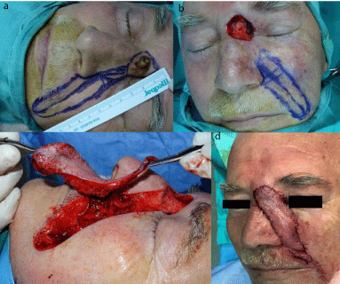
A 66-year-old man presented with a 1-year history of a ulcerating black mass over the left medial canthal region and bridge of the nose. The tumor measured 18 mm (width) × 24 mm (length) at the first examination. A punch biopsy revealed that the tumor was in fact a basal cell carcinoma. We excised the tumor with a 6 mm safety margin keeping the pericondrial and periosteal layer intact (Figure 1a). The defect included the areas immediately neighboring the medial canthal region and base of the nose (Figure 1b). A propeller nasolabial perforator flap was planned for resurfacing the defect. A 9 cm (length) x 2 cm (width) flap was designed over the nasolabial sulcus and nasal sidewall-cheek junction (Figure 1c). While raising the flap two different vessel bundles were identified and dissected from the surrounding soft tissue-muscle units for tension-free rotation (Figure 1d). After a brief discussion among the team both of the vascular pedicles were kept intact. After meticulous dissection flap was rotated 180 degrees to the defect site and half of the flap is used for coverage of the donor site defect (Figure 1d). The residual lower part of donor site defect was closed primarily and the donor scar was left over the nasolabial sulcus (Figure 1e). After completion of the surgery the capillary refill over the flap was 1,5 secs and no immediate venous problem was noted (Figure 1f). Over the 24 hours following surgery the venous insufficiency ensued and became evident (Figure 2a). Even though couple of stiches were removed over the distal part to release the swelling and to ease the tension it was no use the flap became a dusky purple color and lost its capillary refill after roughly 36 hours (Figure 2b). The flap was deemed as a failure and patient was discharged for a later debridement and possible graft coverage. The patient was recalled after one week for a follow-up control and flap was discovered to regain normal refill apart from the upper 10% percent, which is the marginal segment (Figure 2c). Only superficial de-epithelialization on the most distal part was present and local antibiotic ointment was continued for the duration of weekly followup controls. Swelling was subsided quickly and distal part healed completely after 4 weeks without any additional complications (Figure 2d). The excised tissue margin was histopathologically free of tumor cells. At 6 months postoperatively, no tumor recurrence or deformity was evident.

    Figure 1: The tumor with a 6-mm safety margin keeping the pericondrial and
periosteal layer intact.

    Figure 2: Meticulous dissection flap was rotated 180 degrees to the defect
site and half of the flap is used for coverage of the donor site defect.
Discussion
The nasolabial flap is widely used in facial reconstruction, due to its ease and, reliability. Its use is well known for reconstruction of nasal, cheek defects but its extended indications can be reconstruction of upper lip, anterior floor of the mouth, the lower lip and nasal lining as a turn-over flap [5]. The flap’s rich subdermal plexus confers viability even allowing for a length-base relation of 3:1 instead of 2:1; but in this form its base should ideally measure from 2,5 up to 3,5 cm making the primary closure of the donor site problematic.
It is possible to expand the versatility of nasolabial flap by using its perforator counterpart. The nasolabial perforator flap uses the same donor site as traditional nasolabial flap but implements a freestyle islanded flap nourished by a well-designated vascular pedicle based on angular artery or one of its branches [2]. Flap motion can be either rotation up to full propeller or simple advancement. Once identified, pedicle can be up to dissected up to 3 cm providing necessary mobility to the flap to reach all the way to the base of the nose. By using a designated vascular pedicle we can break free from dimension restrictions such as 3:1 or 2:1 and raise long flaps with a dimension ratio of 9:2 as in this case. An islanded flap can move freely to all areas of mid face and the remaining portion of the flap not covering the defect can fill the donor site making primarily closure possible and easy.
Propeller motion is used in this case utilizing two different vascular pedicles. While the upper pedicle is released just enough to make necessary rotation, the lower pedicle is dissected thoroughly to avoid tension on the pedicle when the lower part of the flap (where the lower pedicle is connected) advanced all the way into the defect site. Propeller motion can become an issue in some cases especially in relatively large flaps. Even though the artery can withstand the twisting motion and the arterial flow can persist, the veins of the perforators can collapse easily. Because of that choosing the right perforator and using the right motion is imperative in survival of the flap. In this particular case, using both of the perforators was probably a mistake. Early transient venous insufficiency is expected with these kind of flaps, but venous outflow problem with this flap was most severe to the point that flap lost all its visible capillary refill after 36 hours and deemed as failure. We think after the 180 rotation the veins of the lower perforator was likely collapsed while the both arteries continued providing robust blood flow to the flap resulting in excessive venous insufficiency. Couple of stiches were taken out for both relieving the tension and interfering with accumulation of blood under the flap causing additional pressure. Despite both this maneuvers, as stated capillary refill was lost over the flap.
Immediate debridement and coverage with graft can be tempting because that way the issue can be resolved quickly and without much fuss. But according to our experience, venous outflow problems with facial perforator flaps are common and can be severe in few instances. We think instead of interfering wait-and-see option should be the way to go because;
1. Almost always the problem resolves in 48-72 hours.
2. These flaps are quiet resilient and can recover from even dire situations.
Having said that, we weren’t expecting full recovery from that point. Our expectation was loss of a significant portion of the flap and having a second surgery after demarcation of the necrosis. There are no reports of a facial perforator flap recovering from this kind of severe venous problem without any intervention, so we thought it would be valuable for presentation.
Conclusion
Free-style perforators on the face are used more and more recently instead of conventional flaps because of their versatility and reliability. The common downside of these flaps are venous insufficiency but it usually resolves in couple of days. We hereby presented a good example for the resilience of the nasolabial flap. Although it is one case and cannot represent a wider scope, we think this recovery shows even in grave situations patience observation and conservative approach can be utilized.
References
- KESIKTAS Erol, Gencel E, Aslaner EE. A useful flap combination in wide and complex defect reconstruction of the medial canthal region: Glabellar rotation and nasolabial VY advancement flaps. Plastic Surgery. 2015; 23: 113-115.
- POSSO Carolina, David Delgado Anaya, Jeison Aguilar Henao, Juan M, Velasquez Gaviria. Nasolabial propeller perforator flap: Anatomical study and case series. Journal of surgical oncology. 2018; 117: 1100-1106.
- GOH, Cindy Siaw-Lin, Joshua Guy Perrett, Manzhi Wong, Bien-Keem Tan. Delayed bipedicled nasolabial flap in facial reconstruction. Archives of plastic surgery. 2018; 45: 253.
- LEE, Jun Yong, Ji Min Kim, Ho Kwon, Sung-No Jung, Hyung Sup Shim, et al. Freestyle local perforator flaps for facial reconstruction. BioMed research international. 2015.
- DURGUN, Mustafa, Elif Sari, Hülda Rifat Özakpinar, Caferi Tayyar Selçuk, et al. The versatile facial artery perforator-based nasolabial flap in midface reconstruction. Journal of Craniofacial Surgery. 2015; 26: 1283-1286.