Special Article – Oral Surgery
Austin J Surg. 2019; 6(20): 1215.
Use of the Intrathoracic Tube for Repositioning Free Flap Pedicle via Transoral Approach
Ye J, Jiang X and Xiao M*
Department of Otolaryngology Head and Neck Surgery, Sir Run Run Shaw Hospital, College of Medicine, Zhejiang University, China
*Corresponding author: Mang Xiao, Department of Otolaryngology Head and Neck Surgery, East Qingchun Road 3. Sir Run Run Shaw Hospital, College of Medicine, Zhejiang University, Hangzhou, 310016. Zhejiang, China
Received: August 13, 2019; Accepted: September 25, 2019; Published: October 02, 2019
Abstract
Background: The common site of oral cavity and oropharyngeal cancer including maxilla, soft palate, tongue, tonsil, buccal mucosa and mouth floor. Oral reconstruction with free flap is always necessary because of surgical resection lead to inevitable functional loss. It has a unique procedure, that is, to reposition the pedicle via transoral approach through tunnel created to recipient vessels. However, the flaps pedicle could be squeezed during the transportation and the tunnel must be wide enough, which may cause extra damages.
Methods: We presented in the current study that intrathoracic tube could be wisely made use of to solve the problem. Patients with oral carcinoma underwent primary surgical resection and neck dissection without mandibulotomy, and immediate reconstruction with radial free forearm flap or superficial fascia layer were appropriate for this technique.
Results: This technique based a modified intrathoracic tube allows the thin and reliable free flaps smoothly transported intraoral, decreasing the need of additional debunking procedures. Time to transport the flap as presented ranged between 3 and 5 minutes. There was no loss of arterial Doppler signal.
Conclusion: We utilized the intrathoracic tube in solving the puzzle of transportation free flap. What’s more, it is the prophase of our design a guidewire-like instrument to “guide” the flap pedicle in intraoral approach. We have already applied for a national medical patent of invention and most oropharyngeal cancer patients could be benefit from it.
Keywords: Oral cavity and oropharyngeal cancer; Intrathoracic tube; Free flap pedicle; Transoral approach
Introduction
Oral cavity and oropharyngeal cancer make up approximately 3.25% of all human cancers and keep on rise in recent years [1]. 90% of them are squamous cell carcinomas. The common site of squamous cell carcinoma of oropharynx including maxilla, soft palate, tongue, tonsil, buccal mucosa and mouth floor. Unlike many cancers, the shifts on treatment of oropharyngeal cancer are focused on non-surgical intervention, as the oropharynx has major function on speech, mastication, swallowing, and aesthetics [2,3]. Nevertheless, for those invasive tumors in advanced stage traditional open surgical approach is still inevitable [2,4]. After the ablative surgery, a reconstruction is always necessary, because defects without reconstruction would lead to inevitable functionless [5]. Immediate radial forearm free flap and anterior-lateral thigh fasia lata free flap are optimal for the oral reconstruction [6-8].
External carotid branches are commonly used for oral microvascular reconstruction [9]. The facial artery and vein are most common recipient vessel for free flap. Superior thyroid artery, arising as the first branch of the external carotid artery, is also commonly used [10]. The advantage of these arteries is obvious when the patient is having a neck dissection because their position in the neck. Namely, the pedicle length is determined by these donor vessel, therefore long pedicle length in facial-oral reconstruction is considerably required [9]. Reconstruction of oral defects has a unique procure in the microvascular surgery, that is, to transport the flap pedicle intraoral in patients through a created tunnel. Generally, the anterior pedicle is firstly clamped to the beaks of hemostats, which goes through the submandibular region, then pulled to the recipient vessel region. The problem is that, the pedicle is easily damaged during the squeezing and stretching, meanwhile, the tunnel must be expanded by the hemosts to make the pedicle through.
We presented in the current study that intrathoracic tube could be wisely made use of to solve the problem without extra damages. This technique allows intraoral transposition of the thin and reliable flap, which are optimal for oral reconstruction, such as fasia lata and radial forearm free flap.
Patients and Methods
Patients with oropharyngeal carcinoma underwent primary surgical resection and neck dissection without mandibulotomy, and immediate reconstruction with radial free forearm flap or superficial fascia layer were appropriate for this technique.
Surgical procedure
Tracheostomy was performed in all patients before surgical resection.
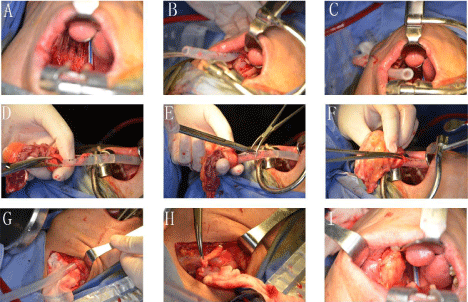
Step 1 - The intrathoracic tube (Size-F34, Diameter=11.33mm) as presented in (Figure 1) is inserted from neck-site through the tunnel approach to oropharynx (Figure 2A).

Figure 1: Schematic illustration of preparing the intrathoracic tube.

Figure 2: Use of the intrathoracic tube as the guidewire in repositioning free pedicle via transoral approach.
Step 2 - Use the hemoststs to grasp the proximal end of tube and gently pull it out. Use a clap to infibulate the distant end of tube (Figure 2B-C).
Step 3 - Cut a tear in about 1cm long at the proximal end of tube, in order to make large opening to contain flap. One hand keeps the tear opening with stiff membrane scissors, one hand put the pedicle of flap into tube, which already injected with saline (Figure 2D-E). Slowly squeeze the pedicle forward into the tube.
Step 4 - Move back scissors to close the tear, so the pedicle could be fixed in the tube (Figure 2F).
Step 5 - Stretch the tube backward slowly until the pedicle is guided to the neck site (Figure 2G). Withdraw the tube smoothly (Figure 2H-I).
Results
We have applied this technique in 10 patients. Mean final flap thickness was 0.7cm. Mean pedicle length was 7.5cm (range from 6 to 9cm). It decreased the need of additional debulking procedures. Time to transport the flap as presented in Figure 2 (A-I) ranged between 3 and 5 minutes. There was no loss of arterial Doppler signal.
Discussion
We introduced a simple and new technique for transposition of free flap into the oral cavity, based on different application of the intrathoracic tube. It reduced the complication of pedicle tear and avoided over-expanded tunnel. Undoubtedly, improving the technique is responsible for the success of oropharyngeal reconstruction. It is prophase of our work, to design a guidewire-like instrument to “guide” the flap pedicle through the tunnel. There is always no ending in proving surgical instrument, an ingenious design makes surgeon pleasing and effective. In another words, sometimes ordinary used in non-routine way could make inspiration.
Funding Sources
This study is sponsored by grants from Medical Health Science and Technology Project of Zhejiang Provincial Health Commission (Grant No.2019336033). Medical Health Science Project of Hangzhou (Grant No. OO20190775).
References
- Mehanna H, Beech T, Nicholson T, El- Hairy I, Paleri V, Robert S, et al. Prevalence of human papillomavirus in oropharyngeal and nonoropharyngeal head and neck cancer--systematic review and meta-analysis of trends by time and region. Head Neck. 2013; 35: 747-755.
- Sims JR, Moore EJ. Primary surgical management with radial forearm free flap reconstruction in T4 oropharyngeal cancer: Complications and functional outcomes. Am J Otolaryngol. 2018; 39: 116-121.
- Zuydam AC, Rogers SN, Brown JS, Vaughan ED, Magennis P. Swallowing rehabilitation after oro-pharyngeal resection for squamous cell carcinoma. Br J Oral Maxillofac Surg. 2000; 38: 513-518.
- L’Heureux-Lebeau B, Odobescu A, Harris PG, Guertin L, Danino AM. Chimaeric subscapular system free flap for complex oro-facial defects. J Plast Reconstr Aesthet Surg. 2013; 66: 900-905.
- Antohi N, Tibirna G, Suharski I, Huian C, Nae S, Stan V, et al. Gastro-omental free flap in oro/hypopharyngeal reconstruction after enlarged ablative surgery for advanced stage cancer. Chirurgia (Bucur). 2013; 108: 503-508.
- Tantawy SA, Kamel DM, Abdelbasset WK, Elgohary HM. Effects of a proposed physical activity and diet control to manage constipation in middleaged obese women. Diabetes Metab Syndr Obes. 2017; 10: 513-519.
- Jeong HH, Hong JP, Suh HS. Thin elevation: A technique for achieving thin perforator flaps. Arch Plast Surg. 2018; 45: 304-313.
- Kerr RP, Hanick A, Fritz MA. Fascia Lata Free Flap Reconstruction of Limited Hard Palate Defects. Cureus. 2018; 10: e2356.
- Haffey TM, McBride JM, Fritz MA. Use of angular vessels in head and neck free-tissue transfer: a comprehensive preclinical evaluation. JAMA Facial Plast Surg. 2014; 16: 348-351.
- Mulholland S, Boyd JB, McCabe S, Rotstein L, Brown D, Yoo J, et al. Recipient vessels in head and neck microsurgery: radiation effect and vessel access. Plast Reconstr Surg. 1993; 92: 628-632.