
Special Article – Surgery Case Reports
Austin J Surg. 2019; 6(21): 1219.
Cholecystocutaneous Fistula after Cholecystectomy
Ping C1, Ping H1, Gang Z1, Xiaoming S2, Kaixiong T2 and Jinxiang Z1*
¹Department of Emergency Surgery, Union Hospital, Tongji Medical College, Huazhong University of Science and Technology, China
²Department of Gastrointestinal Surgery, Union Hospital, Tongji Medical College, Huazhong University of Science and Technology, China
*Corresponding author: Jinxiang Zhang, Department of Emergency Surgery, Union Hospital, Tongji Medical College, Huazhong University of Science and Technology, 1211 Jiefang Avenue, Wuhan, Hubei Province, People’s Republic of China
Received: September 03, 2019; Accepted: October 21, 2019; Published: October 28, 2019
Abstract
A 62 year-old man went to the emergency department with intermittent pain in the right upper anterior abdomen for 7 years and worsening in 10 days. In addition, he had an open cystectomy 48 years ago. On examination, the discharging sinus was at the old surgical scar with pale green liquid secreted. He was diagnosed with cholecystocutaneous fistula post total cholecystectomy. Then he accepted a laparoscopic residual-cholecystectomy and was discharged with clinical cured.
There are few reports reported about cholecystocutaneous fistula after total cholecystectomy. It indicates, “even though a patient has history of cholecystectomy, the complication of cholecystocutaneous fistula can still occur” and proved that laparoscopic cholecystectomy maybe a workable treatment. Any of patient’s present condition and past status should be fully considered to avoid misdiagnosis.
Keywords: Cholecystocutaneous fistula; Laparoscopic cholecystectomy; Cholecystectomy
Case Presentation
A 62 year-old man went to the Department of Emergency Surgery with intermittent pain in the right upper abdomen for 7 years and worsening in 10 days. During the time, the pain in the right upper abdomen was aggravated, and the discharging sinus was formed at the old surgical scar with a pale green liquid flow out. The patient has no symptoms of pyrexia, nausea, vomiting, dizziness, palpitation, radiation pain, etc. 48 years ago when he was only 15 year-old, a diagnosed as acute suppurative cholecystitis was made since he had a history of 2 years recurrent right upper abdominal pain and 1 week of aggravation with systemic fever and swelling of the right upper abdominal wall. Then he accepted an emergency open exploration surgery with a total cholecystectomy in Sep 23rd 1970, the 2nd day after his admission.
On physical examination, patient was conscious, cooperative and well oriented. His pulse, body temperature and blood pressure were normal. On local examination, a discharging sinus was seen in the right upper abdomen through old surgical scar. Pale green liquid was flowing after pressing around it.
Experimental examination: The white cell count was normal. Liver function tests were normal except for a slight increase in bilirubin (TBil: 27.3 umol/L).
Ultrasound imaging examination
The original incision subcutaneous skin damage and communication with the abdominal cavity, considering the formation of sinus (Figure 1).
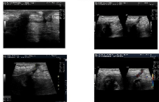
    Figure 1: Ultrasound imaging examination show the original incision
subcutaneous skin damage and communication with the abdominal cavity.
Angiography
The abdominal wall forms a fistula with a depth of 12.2 mm and a width of 2.3 mm. It communicates with the end of the enlarged residual cystic duct, and the contrast agent can reach the duodenum. The filling defect, referred as gallstones, could be found in the residual cystic duct (Figures 2 & 3).
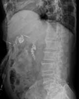
Figure 2: Angiography show the abdominal wall forms a fistula (Lateral).
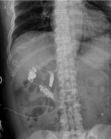
Figure 3: Angiography show the abdominal wall forms a fistula (Positive).
MRCP
The gallbladder is absently, the residual cystic duct was enlarged, and some filling defects were found there; there was no obvious expansion and filling defects in the common bile duct (Figure 4).
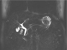
    Figure 4: MRCP: The gallbladder is absently, the residual cystic duct was
enlarged, and some filling defects (stone) were found.
Treatment & Diagnosis
Based on the history and examination, diagnosis of cholecystocutaneous fistula post cholecystectomy was made. After a full routine preoperation examination, a laparoscopic exploration was carried on. During the operation, the residual gallbladder or cystic duct was found and the volume was about 3cm*2cm, which adhered to the surrounding tissues and parts of intestinal walls. A biliary probe was used to explore the discharging sinus from the outside and it could went directly into the syst duct. The intact fistula through the skin was absolutely dissected out. The adhesion tissue around the rest of the gallbladder or cyst duct was carefully separated. Then the triangle was carefully dissected, the branch of the hepatic artery and the cystic duct were identified confirmed and clamped respectively followed by removal of the residual gallbladder or cyst duct (Figures 5-7). Antibiotics and supportive treatment were given after surgery as routine.
Postoperative pathology
The size of the removed tissue is about 3.8*2.5cm. Its surface is rough and irregular and the structure is incomplete. There are two gallstones visible inside. The thickness of the wall is 0.3-0.7cm. Another gray-white tissue, about 1cm*0.9cm*0.3cm in size, has a sinus in the tissue section. Pathological diagnosis: 1. Chronic residual cholecystitis with gallstones (Figure 5). 2. Sinus is formed with proliferating fibrous tissue (Figure 6).
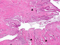
    Figure 5: Histology showing lots of inflammatory cell in the cystic duct. There
is some gland (⁎) which is only can be seen in cystic duct and its suggests
that the residual tissue is the cystic duct.
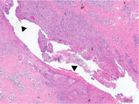
    Figure 6: Histology showing lots of inflammatory cell in gray-white tissue. The
sinus (⁎) can be seen in the section.
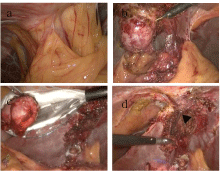
    Figure 7: a: Laparoscopically, extensive adhesions formed the sinus on the
abdominal wall. b: After separation of the adhesion, the residual gallbladder
or cyst duct is visible. c: Removing the residual gallbladder or cyst duct. d:
Explore sinus with cholesterol exploration (⁎).
Discussion
With the use of antibiotics and advances in surgical techniques, spontaneous rupture of the gall bladder into the skin is an extremely rare complication of cholelithiasis in the present era. There are few cases about cholecystocutaneous fistula after abdominal surgery such as gastrectomy, liver abscess surgery, acute cholecystitis drainage [1] and others [2-4]. However, there is only one case reported after subtotal cholecystectomy [5]. What’s more, no literature has reported the occurrence of cholecystocutaneous fistula after total cholecystectomy.
The management of cholecystocutaneous fistula requires initial drainage of any associated abscess and administration of appropriate antibiotics [6]. For a non-cancerous lesion an elective cholecystectomy should be performed when the patient’s general condition is optimized. Moreover, it has been reported that the cholecystocutaneous fistula could be repaired by laparoscopic methods [7]. At present, minimally invasive surgery for non-malignant diseases is the standard procedure for the treatment of many diseases. For patients diagnosed with cholecystocutaneous fistula, it is recommended as far as possible to prefer laparoscopy for minimal trauma, clearer vision. This case indicates that the complication of cholecystocutaneous fistula can still occur although a patient has a history of total cholecystectomy. For post cholecystetomy cholecystocutaneous fistula a full laparoscopic operation is safe and workable.
Acknowledgement
Thanks to the Department of Radiology, Ultrasound, and Pathology of Union Hospital for providing high quality imaging materials and providing guidance for imaging diagnosis.
References
- Ioannidis O, Paraskevas G, Kotronis A, Chatzopoulos S, Konstantara A, Papadimitriou N, et al. Spontaneous cholecystocutaneous fistula draining from an abdominal scar from previous surgical drainage. Ann Ital Chir. 2012; 83: 67-69.
- Surya M, Soni P, Nimkar K. Spontaneous Cholecysto-Cutaneous Fistula Draining Through an Old Abdominal Surgical Scar. Pol J Radiol. 2016; 81: 498-501.
- Dixon S, Sharma M, Holtham S. Cholecystocutaneous fistula: an unusual complication of a para-umbilical hernia repair. BMJ Case Rep. 2014.
- Hawari M, Wemyss-Holden S, Parry GW. Recurrent chest wall abscesses overlying a pneumonectomy scar: an unusual presentation of a cholecystocutaneous fistula. Interact Cardiovasc Thorac Surg. 2010; 10: 828-829.
- Maynard W, McGlone ER, Deguara J. Unusual aetiology of abdominal wall abscess: cholecystocutaneous fistula presenting 20 years after open subtotal cholecystectomy. BMJ Case Rep. 2016.
- Cheng HT, Wu CI, Hsu YC. Spontaneous cholecystocutaneous fistula managed with percutaneous transhepatic gallbladder drainage. Am Surg. 2011; 77: E285-286.
- Malik AH, Nadeem M, Ockrim J. Complete laparoscopic management of cholecystocutaneous fistula. Ulster Med J. 2007; 76: 166-167.