
Special Article – Surgery Case Reports
Austin J Surg. 2019; 6(23): 1223.
Increasing Width of Attached Gingiva by Envelope Technique with Trap Door Technique for Subepithelial Connective Tissue Graft
Trivedi A*
Nr Veeranjali Garden, India
*Corresponding author: Trivedi A, J 5 Staff Quarters, Civil Campus, Nr Veeranjali Garden, Asarwa, Ahmedabad – 380016, Gujarat, India
Received: September 13, 2019; Accepted: October 24, 2019; Published: October 31, 2019
Abstract
Each periodontal plastic & esthetic procedure can be performed using a variety of surgical techniques that are selected based on their advantages and disadvantages relative to the specific clinical presentation of the defect. The present paper focuses on improvement of gingival status by increasing width of attached gingiva by “Envelope technique” with illustrated surgical steps by case presentation.
Keywords: Width of attached gingiva; Envelope technique; Subepithelial connective tissue graft
Introduction
Miller (1993) proposed that the term periodontal plastic surgery is defined as “surgical procedures performed to prevent or correct anatomic, developmental, traumatic or disease induced defects of the gingiva, alveolar mucosa or bone” (Proceedings of the World Workshop in Periodontics 1996) [1].
Miller’s classification of periodontal recession (1985) [2]
Class-I: The recession does not extend to the muco-gingival junction and is not associated with an interdental bone resorption.
There is no pocket and the proximal septa are within normal limits. A root coverage of 100% can be obtained.
Class-II: The recession extends beyond the muco-gingival junction with no interdental bone resorption. There is a rupture of the attachment. A 100% root coverage may also be obtained.
Class-III: The recession is associated with an interdental proximal bone resorption and an extrusion with proximal root exposition.
Root coverage of 100% may not be obtained.
Class-IV: There is proximal bone resorption and the dental papilla is at the same level as the recession. 100% root coverage is impossible.
However, Azzi and Etienne (1998) have shown that root coverage of class III and class IV could be possible.
With the use of the envelope technique [3,4], an envelope is prepared apically and laterally to the recession, the placement of the connective tissue graft is done and to allow for the coronal advancement of the mucosal flap at time of suturing. The sub epithelial connective tissue graft is harvested from the palate or the retromolar pad by the use of a “trap door” approach. Compared to the epithelialized graft, the connective tissue graft is preferable due to less invasive palatal wound and improved esthetic result [5-7].
Case Report
A 48 -year- old male patient reported to Department of Periodontia, with chief complaint of lowering of gum & hypersensitivity in lower left canine since 3 years.
In intraoral soft tissue examination, according to Miller’s classification Grade – II recession was present in relation to lower left canine. Width of attached gingiva was inadequate in lower left canine region (Figure 1).
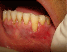
Figure 1: Pre-Operative view.
Surgical technique
Step-1: The recipient site is prepared by first eliminating the sulcular epithelium by an internal beveled incision.
Step-2: Secondly, an envelope is prepared apically and laterally of the recession by split incisions. The depth of the preparation should be 3-5mm in all directions. In apical direction, the preparation of the site should extend beyond the mucogingival junction to facilitate the placement of the connective tissue graft and to allow for the coronal advancement of the mucosal flap at time of suturing.
Step-3: The connective tissue graft was harvested from the palate, following a “trap-door” flap design. A No. 15 blade was used to make a partial-thickness horizontal incision, with a bevel about 3mm apical to the gingival margin of the first premolar, extending towards the first molar. Two vertical incisions were made mesially and distally (Figure 2).
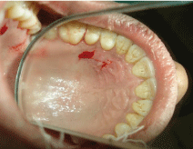
Figure 2: Donor site Preparation: palatal graft is outlined.
Step-4: Tissue forceps was used to lift the prepared palatal flap edge. It was reflected toward the center of the palate and the underlying connective tissue was exposed (Figure 3).
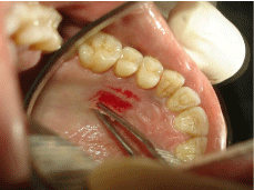
Figure 3: Underlying connective tissue graft was exposed.
Step-5: An incision perpendicular to the bone was made around the edge of the connective tissue, facilitating its reflection from the bone. A small periosteal elevator and knife were used to reflect the connective tissue and harvest it (Figures 4 and 5).
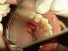
Figure 4: Overall survival, autologous stem cell transplant (ASCT) versus no ASCT (p=0.12).
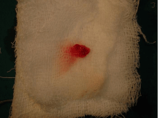
Figure 5: Overall survival, autologous stem cell transplant (ASCT) versus no ASCT (p=0.12).
Step-6: After harvesting the graft, the palatal donor site wound was closed using 4-0 black silk sutures & application of a periodontal dressing is done & Acrylic removable palatal plate for retention of periodontal
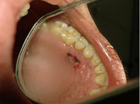
Figure 6: Sutures at donor site.
Step-7: The graft, which is obtained by the trap door approach described above, is inserted into the prepared envelope and positioned to cover the exposed root surface.
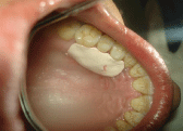
Figure 7: Periodontal dressing was given to protect the donor site.
Step-8: 5-0 polyglactin 910 resorbable Sutures are placed to secure the graft position. A crossed sling sutures had been placed after coronally advanced the mucosal flap. Pressure was applied for 5minutes to closely adapt the graft to the root surface (Figure 9).
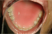
Figure 8: Acrylic removable palatal plate for retention of periodontal pack.
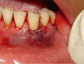
Figure 9: Sutures in place.
Follow up was scheduled on day 3,7,17 & after 16 weeks (Figure 10).
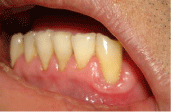
Figure 10: Post-Operative view after 4 months.
Discussion
Short term clinical trials comparing the treatment outcome of the two modalities of free soft tissue graft (Sbordone et al. 1988, Daniel & Cheru 1990, Jahnke et al 1993) have shown that the connective tissue graft results in superior root coverage compared to the epithelialized free soft tissue graft. The color match of connective tissue grafted area to the adjacent gingiva is esthetically also more favorable with the connective tissue graft than that of epithelialized graft [8,9].
In present case, report subepithelial connective tissue graft was taken from the palate, to get superior recession coverage result & for better colour match.
Decision aid model for root coverage surgery (Class I and II defects) (Bouchard 2001) suggest deep wide defect greater than 5mm in size should be best treated by connective tissue graft with envelope technique [10-13].
In present case report to maintain adequate width of attached gingiva envelope technique was performed.
Zabalegui et al 1999 [14] suggests in envelope technique sutures are placed to secure the graft position.
In present case report, crossed sling sutures were placed to coronally advance the mucosal flap and secure the graft.
After a perfect root surface preparation, these techniques are principally based on precise incisions, a delicate manipulation and sutures that permit the stabilization of tissues. Therefore, a better intimacy between the receiving bed and the covering tissues is obtained and results in a thin and stable clot, which will assure a better healing.
Conclusion
The choice of a surgical technique to increase the width of attached gingiva will depend on the etiology of the recession, the position of the tooth and the precise evaluation of the quantity (height) and the quality (thickness) of the apical and lateral gingival tissues adjacent to the recession.
References
- Newman, Takei, Klokkevold, Carranza. Carranza’s Clinical Periodontology, Tenth Edition.
- Miller PD. Classification of marginal tissue recession. Int J Periodont Rest Dent. 1985; 5: 8-13.
- Raetzke PB. Covering localized areas of root exposure employing the “envelope” technique. J Periodontol. 1985; 56: 387-402.
- Allen AL. Use of the supraperiosteal envelope in soft tissue grafting for root coverage. I. Rationale and technique. Int J Periodontics Restorative Dent. 1994; 14: 216-227.
- Edel A. Clinical evaluation of free connective tissue grafts used to increase the width of keratinized gingiva. J Clin Periodontol. 1974; 1: 185-196.
- Langer B, Langer L. Subepithelial connective tissue graft technique for root coverage. J Periodontol. 1985; 56: 715-720.
- Lorenzana ER, Allen EP. The single-incision palatal harvest technique: a strategy for esthetics and patient comfort. Int J Periodontics Restorative Dent. 2000; 20: 297-305.
- Reiser GM, Bruno JF, Mahan PE, Larkin LH. The subepithelial connective tissue graft palatal donor site: anatomic considerations for surgeons. Int J Periodontics Restorative Dent. 1996; 16: 130-137.
- Sullivan HC, Atkins JH. Free autogenous gingival grafts. 3. Utilization of grafts in the treatment of gingival recession. Periodontics. 1968; 6: 152-160.
- Allen AL. Use of the supraperiosteal envelope in soft tissue grafting for root coverage. II. Clinical results. Int J Periodontics Restorative Dent. 1994; 14: 302-315.
- Jepsen K, Heinz B, Halben JH, Jepsen S. Treatment of gingival recession with titanium reinforced barrier membranes versus connective tissue grafts. J Periodontol. 1998; 69: 383-391.
- Levine RA. Covering denuded maxillary root surfaces with the subepithelial connective tissue graft. Compendium. 1991; 12: 568-572.
- Muller HP, Stahl M, Eger T. Root coverage employing an envelope technique or guided tissue regeneration with a bioabsorbable membrane. J Periodontol. 1999; 70: 743-751.
- Tal H, Moses O, Zohar R, Meir H, Nemcovsky C. Root coverage of advanced gingival recession: A comparative study between acellular dermal matrix allograft and subepithelial connective tissue grafts. J Periodontol. 2002; 73: 1405-1411.
- Zabalegui I, Sicilia A, Cambra J, Gil J, Sanz M. Treatment of multiple recessions with the tunnel subepithelial connective tissue graft: A clinical report. Int J Periodontics Restorative Dent. 1999; 19: 199-206.