Special Article – Laparoscopic Surgery
Austin J Surg. 2019; 6(26): 1233.
Transvaginal Extraction of Specimens in Totally Laparoscopic Sigmoidectomy or Anterior Resection Combined with Hysterectomy and Bilateral Adnexectomy for Locally Advanced Colorectal Cancer
Yu S, Deng J, Cao J, Luo T, Yong Ji* and Zhen Z*
Department of General Surgery, The First People’s Hospital of Foshan (Foshan Hospital of Sun Yat-sen University), China
*Corresponding author: Yong Ji and Zuojun Zhen, NO.81 Lingnan Road North, The Department of General Surgery, First People’s Hospital of Foshan (Foshan Hospital of Sun Yat-sen University), Foshan 528000, China
Received: October 14, 2019; Accepted: December 03, 2019; Published: December 10, 2019
Abstract
Purpose: Sigmoid colon cancer or rectal cancer that involves the uterus and ovary is common in clinical practice, and treatment usually requires removal of the two organs. In an era of minimally invasive surgery, totally laparoscopic sigmoidectomy or anterior resection combined with hysterectomy and bilateral adnexectomy is an encouraging procedure.
Objective: This study aimed to determine the feasibility, safety, technique and short- and long-term outcomes of transvaginal extraction of specimens in totally laparoscopic sigmoidectomy or anterior resection combined with hysterectomy and bilateral adnexectomy for locally advanced colorectal cancer.
Methods: From January 2000 to December 2014, consecutive patients with sigmoid colonic or rectal cancer which had locally invaded the uterus and ovary underwent totally laparoscopic sigmoidectomy or anterior resection combined with hysterectomy and bilateral adnexectomy. The specimens were extracted via the vagina.
Results: For the 36 patients, none required conversion to open laparotomy. The 90-day operative mortality rate was 0%. The mean operative time was 173±13 minutes. The mean intraoperative blood loss was 80±12 ml. The mean postoperative VAS score on postoperative day 1 was 2.9±1.0, and no patients required painkillers after the operation. All patients were able to get out of bed on day 1 of surgery. The mean time to pass the first flatus was 82±18 hours after surgery. The mean postoperative hospital stay was 6.9±1.8 days. The postoperative complication rate was 31% (11/36), which included anastomoticcutaneous fistula (n=1), vaginal-cutaneous fistula (n=1), rectovaginal fistula (n=2), early postoperative adhesive intestinal obstruction (n=3), acute urinary retention (n=2), and pulmonary infection (n=2). The median follow-up was 62 (range 15~128) months. The median overall survival was 64.5 months. The 1-, 3- and 5-year overall survival rates were 100%, 81% and 61%, respectively.
Conclusions: Transvaginal extraction of specimens in totally laparoscopic sigmoidectomy or anterior resection combined with hysterectomy and bilateral adnexectomy for locally advanced colorectal cancer was technically feasible and safe. It had the advantages of minimal invasiveness with quick recovery. The long-term follow-up oncological outcomes were good.
Keywords: Transvaginal extraction of specimens; Colorectal cancer; Totally laparoscopic surgery; Hysterectomy and bilateral adnexectomy
Introduction
Locally advanced sigmoid colonic or rectal cancer with invasion of uterus or ovary but without distant metastases is not rare. Treatment requires en bloc resection of the two organs. In this modern era of minimally invasive surgery, either laparoscopic sigmoidectomy/ anterior resection or hysterectomy with bilateral adnexectomy are commonly performed surgical procedures [1-3]. However, laparoscopic en bloc resection of these two adjacent organs have rarely been reported [4], and there have been virtually no reports on transvaginal extraction of such specimens. For more than ten years, patients with sigmoid colonic or rectal cancer with local invasion of uterus and ovary underwent totally laparoscopic sigmoidectomy or anterior resection combined with hysterectomy and bilateral adnexectomy in our center, and the specimens were extracted via the vagina.
Data and Methods
Clinical data
This is a retrospective study on prospectively collected data on consecutive married women who underwent totally laparoscopic sigmioidectomy or anterior resection combined with hysterectomy and bilateral adnexectomy for locally advanced colorectal cancer.
All patients presented with bloody or mucous stools and underwent preoperative colonoscopy with biopsy showing well to moderately differentiated adenocarcinoma. CT or MRI was done to assess tumor resectability and to rule out distant metastases. All patients had tumors adherent to the uterus or ovary and were determined by gynecologists to require total hysterectomy and bilateral adnexectomy to achieve en bloc resection of the colorectal cancer. The surgical procedures were approved by the Ethics Committee of our Hospital, and all the operations were carried out in accordance with the relevant guidelines and regulations as stipulated by this Committee. Before operation, all patients underwent neoadjuvant concurrent radiochemotherapy consisting of DT 50Gy in 25 fractions over 5 weeks, concurrent with 4 cycles of chemotherapy [each cycle consisting of 14 days of Xeloda (1000mg/m2, bid) followed by seven days off]. After operation, all patients underwent 4-6 cycles of adjuvant chemotherapy using XELOX [5]. All patients gave informed consent for the operations and for their data to be used for research purposes.
Surgical procedures
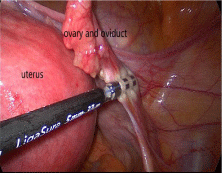
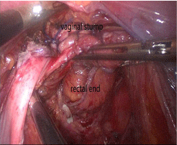
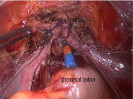
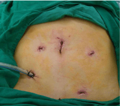
The patient was put under general anesthesia with tracheal intubation, and placed in a lithotomy position. An indwelling urinary catheter was inserted, followed by vaginal douching. The operation was carried out using a five-port technique (Figure 1). After CO2 pneumoperitoneum was established, pressure was maintained at 12 mmHg. Routine intraperitoneal exploration was performed to determine tumor positions, sizes and involvements, and feasibility of transvaginal specimen extraction. The sigmoid mesentery was freed at the root with an ultrasonic scalpel (Harmonic, Johnson & Johnson, USA). The origins of the inferior mesenteric artery and vein were dissected. The left ureter was protected. The descending and sigmoid colon and the posterior wall of the rectum were dissected in the Toldt’s plane to the scheduled transection site. The sigmoid mesocolon was trimmed and the marginal vascular arcade was protected. Gynecologists then started mobilization of the uterus. The course of the left ureter was identified. Ligasure (Valleylab, USA) was used to resect the ligaments of the funnel pelvis and the round ligaments (Figure 2). The vesical peritoneal reflection was incised, and the bladder was pushed downward. An assistant uplifted the patient’s uterus via the vagina with a uterine manipulator. An ultrasonic scalpel was then used to incise the anterior fornix of the vagina followed by extension of the incision bilaterally. Bilateral blood vessels and ligaments were divided and the incision around the posterior vaginal fornix of the uterus was completed. The uterus and bilateral accessory structures were freed. The vaginal stump was not closed, and a gauze pad wrapped in a sterile rubber glove was used as a vaginal plug to prevent gas leakage. The front wall of the rectum was fully dissected. At the scheduled transection site, the wall of the rectum was skeletonized, and a stapler (Echelon 60, Ethicon Endo- Surgery, Cincinnati, USA) was used to cut and close the rectum at the distal end 5 cm away from the tumor. If the sigmoid colon was long enough, the specimen was put into a plastic bag and was retrieved transvaginally. The colon was dissected at the proximal end about 10 cm away from the tumor. A stapling anvil was put onto the divided end of the proximal colon and anchored with a purse-string stitch. The proximal colon was placed transvaginally back to the intraperitoneal cavity. If the sigmoid colon was too short for transvaginal sigmoid colonic resection, a pair of bowel forceps was used to clamp the upper sigmoid colon to prevent spillage of colonic contents. The colon was transected at the proximal end 10 cm away from the tumor. The specimen which included the rectosigmoid colon combined with the uterus and its bilateral appendages were put into a disinfected plastic bag and retrieved via the vagina. A stapling anvil was introduced through the vagina and placed into the cut end of the proximal colon and anchored with a purse–string suture under laparoscopic vision. The vaginal stump was closed using a 2-0 absorbable suture under laparoscopic vision (Figure 3). The pelvic cavity was irrigated with sterile saline. Finally, a stapler (CDH28, Covidien, USA) was inserted through the anus, and colon-rectal anastomosis was completed under laparoscopic vision (Figure 4). A double-lumen drainage tube was inserted through the primary port site at the McBurney’s point and placed in the pelvic cavity of the patient (Figure 5). The en bloc resection specimen of the colorectum combined with the uterus and its bilateral appendages, and the mesenteric lymph nodes were studied histopathologically.

    Figure 1: Port sites for the operation. A – 12 mm port for the laparoscope.
B – 12 mm port for the surgeon’s operative port. C, D, E – 5 mm ports for the
surgeon and assistants’ axillary ports.

    Figure 2: Ligasure was used to resect bilateral pelvic funnel ligaments and
round ligaments.

    Figure 3: The rectum was cut at the distal end 5 cm away from tumor by a
stapler (Echelon 60, Ethicon Endo-Surgery, Cincinnati, USA). The vaginal
stump was closed using 2-0 absorbable suture under laparoscopic vision.

    Figure 4: Colon-rectal anastomosis was completed by a stapler (CDH28,
Covidien, USA).

    Figure 5: A double-lumen drainage tube was placed in the pelvic cavity and
retrieved from the primary operative port via the McBurney’s point.
Follow-up
The patients were regularly followed-up in our outpatient’s clinic. If they failed to attend the follow-up visits, they were contacted by our research nurse by phone calls to update on their health conditions.
Results
There were 36 married female patients. The mean age was 60.6 years (range 44 – 76). The 90-day surgical mortality rate was 0%. The mean operative time was 173±13 minutes. The mean intraoperative blood loss was 80±12 ml. There was no conversion to open surgery. Postoperative pain was evaluated by the Visual Analogue Score (VAS) [6], and the mean VAS on postoperative day 1 was 2.9±1.0. No patients needed painkillers after the operation. All patients were able to get out of bed on the first day after operation. The mean time to passage of the first flatus was 82±18 hours. The mean postoperative hospital stay was 6.9±1.8 days.
The overall postoperative complication rate was 31% (11/36), which included anastomotic-cutaneous fistula (n=1), vaginalcutaneous fistula (n=1), rectovaginal fistula (n=2), early postoperative adhesive intestinal obstruction (n=3), acute urinary retention (n=2), and pulmonary infection (n=2). All these complications responded to conservative treatment.
All patients had R0 resections on histopathological examination of the resected specimens. The average number of harvested lymph nodes was 14.6 (range 12 to 18). Using the 7th edition of UICC TNM Classification of Malignant Tumours, 15 patients were classified as stage IIc (T4bN0M0, without lymph node metastasis) and 21 patients as stage IIIc (T4bN1-2M0, with lymph node metastasis).
On follow-up, all patients could resume light physical activities. Ten patients who had not reached retirement age continued their fulltime employment work. 19 patients resumed normal sexual activities after surgery, without dyspareunia or other discomfort.
This study was censored on December 31, 2016. At a median follow-up of 62 (range 15~128) months, 24 patients had died of recurrence or metastases, with a median survival of 64.5 months. There was no recurrent or implanted tumor in the vaginal stump. The 1-, 3- and 5-year overall survival rates were 100%, 81%, and 61% respectively.
Discussion
In this era of minimally invasive surgery, laparoscopic sigmoidectomy, or anterior resection has become popular. Studies showed that laparoscopic colorectal resection results in less postoperative pain, earlier recovery, shorter hospital stay, and improved cosmesis without affecting short- and long-term oncological outcomes [7-9]. However, most laparoscopic colorectal cancer surgeries require an incision of about 4-6 cm long in the abdominal wall to retrieve the resected specimens, or in some cases to perform colorectal anastomosis. Such an incision minimizes the advantages of minimally invasive surgery [10-12].
In recent years, some surgeons have showed special interest in the use of laparoscopic surgery combined with natural orifice specimen extraction. As specimens are retrieved without the need to make any incision in the abdominal wall, the combined treatment results in less surgical trauma and significantly reduces the incidence of incisionrelated complications.
In general, specimens are most commonly extracted through the anus or the vagina. The reports on laparoscopic colorectal cancer surgery with specimens extracted through the anus demonstrated good results [13-16].
The vaginal route has been widely used in conventional vaginal surgery and in removal of pelvic masses. In 1993, Delvaux et al first reported on transvaginal extraction of specimens after laparoscopic cholecystectomy [17]; followed later by reports on transvaginal extraction of spleen [18] and kidney [19]. Subsequent reports on transvaginal extraction of specimens after laparoscopic surgery for colorectal cancer demonstrated good short- and long-term results [20-22]. In the present study, 36 patients with locally advanced colorectal cancer with invasion to the uterus or ovary were treated with totally laparoscopic en bloc resection of the colorectal cancer and the uterus and its appendages. The specimens were retrieved through the vagina. A prerequisite for transvaginal extraction of specimens is the presence of a vaginal opening after hysterectomy. As the vagina is distensible, it allows transvaginal extraction of specimens without resulting in rupture of the specimens. This route is better than the transanal route as anorectal injuries can occur by extracting a large specimen through the anus.
To achieve good results of this operation, particular attention should be paid to: (1) strict preoperative intestinal and vaginal preparations. Adequate bowel preparation and cleansing of the vagina with antiseptics are important measures to decrease postoperative sepsis. (2) En bloc resection of the tumor is necessary. Separation of a tumor from the uterus to facilitate tumor resection is unwise, because it may lead to intraperitoneal disseminations. If the patient’s uterus is too large and affects exposure and separation of the rectal anterior wall, an ultrasonic scalpel can be used to cut through the myometrium in the normal uterine wall to separate the tumor-involved uterine wall from the uterine body. (3) All surgical specimens should be extracted in a disinfected plastic bag to prevent tumor implantation and contamination of the vaginal wound. In this study, a sheathshaped sterile bag was used as previously reported by us on extraction of specimens through the anus [18,19]. (4) If the colon is transected intraperitoneally, a pair of bowel forceps should be used to clamp the upper colon tightly to prevent spillage of colonic contents and a gauze pad should be used to protect the surrounding tissues. Sterile saline should be used to irrigate the abdominal and pelvic cavities after specimen retrieval. (5) If the tumor or uterus is too large to be extracted transvaginally, an abdominal wound should be made to take out the specimens. Violent pulling should not be allowed as it would tear the perineum or cause rupture and implantation of tumor cells. We have not encountered this situation in our experience in our 36 patients.
In the present study, 1 patient had a anastomotic-cutaneous fistula and 2 patients had a rectovaginal fistula. Excluding the patient with a vaginal-cutaneous fistula which did not involve the anastomosis, the incidence of anastomotic fistula in this study was 8.3% (3/36). This incidence is similar to the reported incidence of anastomotic fistula after laparoscopic proctectomy [23,24]. The results suggested that hysterectomy and bilateral adnexectomy did not increase the incidence of anastomotic fistula.
One patient had a vaginal stump fistula, cause by imprecise suture closure. To prevent vaginal fistula, particular attention should be paid to: (1) Thorough vaginal preparation before operation to minimize the amount of bacteria in the vagina. (2) Separation of the rectovaginal space should be handled with caution to prevent damage to the vaginal wall. (3) When the vaginal stump is closed using a 2-0 absorbable suture under laparoscopic vision, continuous suturing is preferred with a uniform distance (preferably 0.8 cm) between sutures, and the knot-tying should be tight and reliable. (4) A drainage tube should be placed into the pelvic cavity to prevent local infection resulting in rectovaginal fistula.
In this study, the 1-, 3- and 5-year overall survival rates were 100%, 81%, and 61% respectively. It showed the oncological effect of this minimally invasive surgical procedure was satisfactory. Given the small sample size in this study, the patients were not further divided according to the different pathological stages for further analyses. Further studies using a larger sample size is required for such analyses.
In conclusion, transvaginal extraction of specimens after totally laparoscopic sigmoidectomy or anterior resection combined with hysterectomy and bilateral adnexectomy to treat locally advanced colorectal cancer was safe and feasible. This minimally invasive surgery had good short- and long-term results.
Acknowledgment
We express our sincere thanks to Professor Wan Yee Lau of the Chinese University of Hong Kong. In the writing of this paper, we have received the selfless help from Professor Wan Yee Lau.
Ethical Approval
The study has the approval of the Medical Ethics Committee of the First People’s Hospital of Foshan (Foshan Hospital of Sun Yat-sen University).
References
- Seki Y, Ohue M, Sekimoto M, Takiguchi S, Takemasa I, Ikeda M, et al. Evaluation of the technical difficulty performing laparoscopic resection of a rectosigmoid carcinoma: visceral fat reflects technical difficulty more accurately than body mass index. Surg Endosc. 2007; 21: 929-934.
- Agha A, Benseler V, Hornung M, Gerken M, Iesalnieks I, Fürst A, et al. Longterm oncologic outcome after laparoscopic surgery for rectal cancer. Surg Endosc. 2014; 28: 1119-1125.
- Jung YW, Lee M, Yim GW, Lee SH, Paek JH, Kwon HY, et al. A randomized prospective study of single-port and four-port approaches for hysterectomy in terms of postoperative pain. Surg Endosc. 2011; 25: 2462-2469.
- Daraï E, Ballester M, Chereau E, Coutant C, Rouzier R, Wafo E. Laparoscopic versus laparotomic radical en bloc hysterectomy and colorectal resection for endometriosis. Surg Endosc. 2010; 24: 3060-3067.
- Xie Q, Wen F, Wei YQ, Deng HX, Li Q. Cost analysis of adjuvant therapy with XELOX or FOLFOX4 for colon cancer. Colorectal Dis. 2013; 15: 958-962.
- Kong SK, Onsiong SM, Chiu WK, Li MK. Use of intrathecal morphine for postoperative pain relief after elective laparoscopic colorectal surgery. Anaesthesia. 2002; 57: 1168-1173.
- Day AR, Smith RV, Jourdan IC, Rockall TA. Survival following laparoscopic and open colorectal surgery. Surg Endosc. 2013; 27: 2415-2421.
- Kellokumpu IH, Kairaluoma MI, Nuorva KP, Kautiainen HJ, Jantunen IT. Short- and long-term outcome following laparoscopic versus open resection for carcinoma of the rectum in the multimodal setting. Dis Colon Rectum. 2012; 55: 854-863.
- Kaushik S, Akhter K, Rufford B, Ind TE, Kolomainen DF, Butler J, et al. The use of laparostomy in patients with gynecologic cancer: first report from a UK cancer center. Int J Gynecol Cancer. 2013; 23: 951-955.
- Wilson MZ, Hollenbeak CS, Stewart DB. Laparoscopic colectomy is associated with a lower incidence of postoperative complications than open colectomy: a propensity score-matched cohort analysis. Colorectal Dis. 2014; 16: 382-389.
- Lee SM, Kang SB, Jang JH, Park JS, Hong S, Lee TG, et al. Early rehabilitation versus conventional care after laparoscopic rectal surgery: a prospective, randomized, controlled trial. Surg Endosc. 2013; 27: 3902-3909.
- Owada Y, Okamura S, Murata K. A case report of wound-site recurrence two years after laparoscopic colectomy for colon cancer. Gan To Kagaku Ryoho [Article in Japanese]. 2012; 39: 2273-2274.
- Franklin ME Jr, Liang S, Russek K. Integration of transanal specimen extraction into laparoscopic anterior resection with total mesorectal excision for rectal cancer: a consecutive series of 179 patients. Surg Endosc. 2013; 27: 127-132.
- García JI, Gijón MM, Castro PG, Folgueras AL, González JJ. Laparoscopic recto-sigmoidal resection with transanal extraction of the surgical specimen (NOSE) as a treatment for early colorectal cancer (description of the technique) [Article in Spanish]. Cir Esp. 2011; 89: 547-549.
- Deng J, et al. Application of transanal specimen extraction in laparoscopic high anterior rectal resection for colorectal cancer. Chinese Journal of Gastrointestinal Surgery [Article in Chinese]. 2012; 15: 432.
- Deng J, et al. Application of transanal drawing-resection in laparoscopic anterior resection in high-middle rectal carcinoma. Chinese Journal of Gastrointestinal Surgery [Article in Chinese]. 2014; 17: 88-89.
- Delvaux G, Devroey P, De Waele B, Willems G. Transvaginal removal of gallbladders with large stones after laparoscopic cholecystectomy. Surg Laparosc Endosc. 1993; 3: 307-309.
- Vereczkei A, Illenyi L, Arany A, Szabo Z, Toth L, Horváth OP. Transvaginal extraction of the laparoscopically removed spleen. Surg Endosc. 2003; 17: 157.
- Kishore TA, Shetty A, Balan T, John MM, Iqbal M, Jose J, et al. Laparoscopic donor nephrectomy with transvaginal extraction: initial experience of 30 cases. J Endourol. 2013; 27: 1361-1365.
- Nishimura A, Kawahara M, Honda K, Ootani T, Kakuta T, Kitami C, et al. Totally laparoscopic anterior resection with transvaginal assistance and transvaginal specimen extraction: a technique for natural orifice surgery combined with reduced-port surgery. Surg Endosc. 2013; 27: 4734-4740.
- Diana M, Perretta S, Wall J, Costantino FA, Leroy J, Demartines N, et al. Transvaginal specimen extraction in colorectal surgery: current state of the art. Colorectal Dis. 2011; 13: 104-111.
- Park JS, Choi GS, Lim KH, Jang YS, Kim HJ, Park SY, et al. Clinical outcome of laparoscopic right hemicolectomy with transvaginal resection, anastomosis, and retrieval of specimen. Dis Colon Rectum. 2010; 53: 1473-1479.
- Shearer R, Gale M, Aly OE, Aly EH. Have early postoperative complications from laparoscopic rectal cancer surgery improved over the past 20 years? Colorectal Dis. 2013; 15: 1211-1226.
- Scarpinata R, Aly EH. Does robotic rectal cancer surgery offer improved early postoperative outcomes? Dis Colon Rectum. 2013; 56: 253-262.