Special Article – Surgery Case Reports
Austin J Surg. 2020; 7(1): 1238.
Neuroendoscopic Approach for the Management of a Rare Case of Isolated Third Ventricle Glioblastoma
Belfquih H* and Akhaddar A
Department of Neurosurgery, Avicenne Military Hospital, Morocco and Mohammed V University in Rabat, Morocco
*Corresponding author: Belfquih H, Department of Neurosurgery, Avicenne Military Hospital, 40000, Marrakech, Morocco
Received: December 04, 2019; Accepted: January 02, 2020; Published: January 09, 2020
Abstract
Introduction: Glioblastoma is the most common and the most malignant type of gliomas. Cerebral hemispheres are usual locations for gliomas. Isolated third ventricular presentation is very rare for glioblastomas. Here we presente a new case of isolated third ventricular glioblastoma manged with neuroendoscopic approach.
Case Description: A 8-year-old child was admitted with headache, blurred vision and confusion. MRI had showed third ventricular mass lesion with obstructive hydrocephalus. Neuroendoscopic biopsy was performed and a second ventriculoperitoneal shunt was inserted from the opposite site. Histopathological diagnosis was glioblastoma. Radiotherapy and chemotherapy were started immediately after the surgery. Patient’s hydrocephalus has resolved and she was well at postoperative 6th month.
Discussion and Conclusion: Differential diagnosis of high grade gliomas located in the third ventricle is extremely difficult due to the rarity of these tumours. The presence of necrosis and ring enhancement on MRI, are suggestive signs of glioblastoma, yet not pathognomonic. Tumor histology is crucial to yield the final diagnosis. Radical excision of third ventricle glioblastomas should be avoided for two reasons: the high risk of hypothalamic injury, and the outcome will not be improved. neuroendoscopy is less invasive and effective method for management of these tumors
Keywords: Third ventricle; Glioblastoma; Biopsy; Neuroendoscopy; Hydrocephalus; Ventriculo-peritoneal shunt
Introduction
Glioblastomas are the most frequent brain tumor, accounting for approximately 12-15% of all intracranial neoplasms and 50-60% of all astrocytic tumours [1]. Usual tumor location for glioblastomas is cerebral hemispheres, the presence of this tumor in the third ventricle can be considered exceptional. We present a rare case of isolated third ven- tricular glioblastoma, which has been managed with endoscopic biopsy and adjuvant chemoradiotherapy. We discuss clinical, radiological, therapeutic aspects of this rare pathology, with a literature review.
Case Report
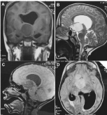
A 8-year-old child was admitted with altered level of consciousness accompanied by headaches, blurred vision, nausea, and vomiting. This was preceded by worsening of short-term memory loss, increasing generalized confusion, increasing somnolence, decreased activity, and cognitive decline for several weeks. MRI study confirmed the presence of a third ventricle lesion with obstructive hydrocephalus. The tumor was predominantly hypointense on T1-weighted and hyperintense on T2-weighted images, with ring enhancement after Gadolonium administration (Figure 1). Neuroendoscopic biopsy was performed and a second ventriculoperitoneal shunt was inserted from the opposite site. Histopathological diagnosis was glioblastoma. The patient’s hydrocephalus has resolved. The patient’s headaches, nausea, and vomiting also improved. The family noted a marked increase in cognition affect and ability postoperatively. Radiotherapy and chemotherapy were started immediately after the surgery and she was well at postoperative 6th month.

    Figure 1: Preoperative magnetic resonance imaging. A: T1-weighted
sequence, coronal section showing a homogenous hypointense solitary
third ventricle mass with hydrocephalus. B: Sagittal T2 - weighted image
of the same lesion wich is hyperintense. C: Contrast-enhancing sagittal Tlweighted
MRI, demonstrating the solitary mide ring enhancing lesion in the
third ventricle. D: Axial FLAIR MRI showing a hyperintense mass at the same
area with active hydrocepalus.
Discussion
Gliomas are the most common primary tumors of CNS in adult patients. Glioblastoma is the most malignant and most common type (50-60%) of all gliomas [2]. Usual tumor location for gliomas is cerebral hemispheres. Diagnosis of a glioblastoma presenting as a unique mass entirely restricted to within the third ventricle cavity can be considered exceptional.
The most probable origin of a third ventricle glioma should be located at any of the structures that surround this cavity, mainly hypothalamic and thalamic nuclei, septum pellucidum, fornices and septal nuclei. Since all these structures were intact on the preoperative MRI studies of our case above mentioned, it could be speculated that the initial tumoral cells would have developed at the subependymal level, breaking early in their growth through the ependymal layer towards the third ventricle cavity [2].
In differential diagnosis list of the tumors presenting in the third ventricle, there are plenty of tumors such as colloid cyst, meningioma, germinoma, craniopharyngioma, lymphoma, choroid plexus papilloma, subependymal giant cell astrocytoma, chiasmatic and hypothalamic benign astrocytoma [3]. Ring enhancement and presence of necrosis on MRI, are suggestive signs of glioblastoma, yet not pathognomonic [4]. Tumor histology is crucial to yield the final diagnosis [5].
Total resection is possible yet hard due to very deep location of the tumor at the center of the brain, within surrounding delicate tissues and fragile vessels. Malignant nature and intractable course of the tumor should be considered while planning treatment. Endoscopic approach to the third ventricle is a minimally invasive procedure compared to conventional craniotomy. By this approach not only access to the tumor itself, but also relief of the hydrocephalus caused by the tumor could be managed simultaneously using ventriculostomy, septostomy or aqueductal stenting [6]. Operative time and morbidity rates attributed to endoscopic approach to third ventricle are lower than microsurgical approach. However, complete resection rate is higher in microsurgical approach. Radical excision of third ventricle glioblastomas should be avoided for two reasons: the high risk of hypothalamic injury, and the outcome will not be improved. neuroendoscopy is less invasive and effective method for management of these tumors [6].
References
- Kleihues P, Louis DN, Scheithauer BW, Rorke LB, Reifenberger G, Burger PC, et al. Glioblastoma. In: Kleihues P, Cavenne WK, editors. WHO tumors of the nervous system. Lyon: IARC Press. 2000; 29-39.
- Prieto R, Pascual JM, Roda JM. Third ventricle glioblastoma. Case report and review of literature. Clin Neurol Neurosurg. 2006; 108: 199-204.
- Lee TT, Manzano GR. Third ventricular glioblastoma multiforme: case report. Neurosurg Rev. 1997; 20: 291-294.
- Deramond H, Pruvo J, Gondry C, Baledent O, Desenclos C. Imaging of tumors of the third ventricle. Neurochirurgie. 2000; 46: 239-256.
- Yilmaz B, EksI MS, Demir MK, Akakin A, Toktas ZO, Yapicier O, et al. Isolated third ventricle glioblastoma . Springer Plus. 2016; 5: 115.
- Schroeder HW. Intraventricular tumors. World Neurosurg. 2013; 79: S17e15- S17e19.