Case Report
Austin J Surg. 2020; 7(3): 1249.
Heterotopic Pancreatic Tissue Mimicking a Submucosal Gastric Tumour
Bigdeli HR1,2, Petrushnko W1, Isaacs A1 and Ghusn M1*>
1Department of Surgery, The Tweed Hospital, Australia
2Griffith University, School of Medicine, Gold Coast, Australia
*Corresponding author: Ghusn M, Upper Gastrointestinal Department, The Tweed Hospital, Tweed Heads, Australia
Received: June 12, 2020; Accepted: July 15, 2020; Published: July 22, 2020
Abstract
Background: Heterotopic pancreatic tissue is a rare entity that was first described in an ileal diverticulum in the 18th century. Clinically, they are a diagnostic dilemma as they can present similar to other gastrointestinal tumors. The majority are asymptomatic, but others can present with inflammation, pain, bleeding, and obstruction depending on their size and location. A review of literature has shown that these heterotopic pancreatic tissues rarely exceed 20mm in size, with the largest previously recorded being 35mm. None to our knowledge were due to a ball-valving mechanism.
Case Report: This case report presents a 22-year-old female with a 6-month history of obstructive symptoms secondary to a heterotopic pancreatic tissue. Due to its large size and location in the gastric antrum, the intermittent obstructive symptoms were a direct result of the ball-valving of the mass. The tissue was excised and subsequently measured to be greater than the largest previously recorded heterotopic pancreas in literature.
Discussion: Although rare, heterotopic pancreatic tissues should be considered as a possible differential diagnosis for gastrointestinal stromal tumours, and if symptomatic, surgical resection should be performed.
Keywords: Heterotopic Pancreas; Gastric Outlet Obstruction; GIST; Gastrectomy
Abbreviations
CT: Computed Tomography; GIST: Gastrointestinal Stromal Tumour
Introduction
Heterotopic, or ectopic pancreas, is a rare entity which is defined as the presence of pancreatic tissue without anatomic and vascular continuity with the main body of the pancreas [1]. It was first described by Schultz in the 18th century in an ileal diverticulum [2]. It may occur in a variety of locations within the gastrointestinal tract, but has a propensity to affect the stomach and small intestine [1]. Clinically, it is a diagnostic challenge as Gastrointestinal Stromal Tumours (GISTs) can present in similar fashion [3]. In most cases, heterotopic pancreatic tissue is asymptomatic, but can become clinically evident when it presents with inflammation, pain or bleeding [4]. Depending on the size and location of the tissue, patients can also present with obstructive symptoms. We present are port of a 22-year-old female with intermittent gastric outlet obstruction secondary to a large heterotopic pancreatic tissue.
Case Report
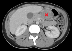
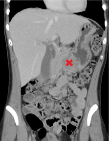
A 22-year-old female with a 6-month history of episodic epigastric pain, nausea and post-prandial vomiting. More recently the episodes had increased in frequency. Investigations including CT were performed. The CT Abdomen showed a 55x42mm submucosal mass in the gastric antrum, with a likely diagnosis of a Gastrointestinal Stromal Tumour (GIST) (Figure 1 and 2). A gastroscopy was later performed and showeda large, submucosal, non-circumferential mass on the anterior wall of the gastric antrum with no bleeding and no stigmata of recent bleeding (Figure 3). A biopsy was taken with the histopathology being non-diagnostic. Given the unclear diagnosis and with the possibility of malignancy, an enblocdistal gastrectomy and D1 nodal dissection was performed one week later. Intraoperatively, the mass was found to have adhered to a small portion of the proximal transverse colon with surrounding palpable lymphadenopathy within the mesentery. The involved section of the transverse colon was resected en bloc. The histopathology demonstrated heterotopic pancreatic tissue (Heinrich type1), with associated active inflammation and fibrosis however no evidence of malignancy. The tissue was measured to be 40x35x40mm, making it the largest recorded heterotopic pancreatic tissue.
Discussion
Although the exact pathogenesis of heterotopic pancreatic tissue has not been identified, there have been two main embryological theories. The first proposes that during the development of the pancreas form several evaginations from the wall of the primitive duodenum, one or more evaginations may remain within bowel wall, which after migrating with the development of the gastrointestinal tract gives rise to the ectopic pancreatic tissue [1]. The second theory suggests that the heterotopic pancreas may be due to its separation from the primitive pancreas during embryonic rotation [5].

Figure 1: Axial view of lesion marked with a red cross.

Figure 2: Coronal view of lesion marked with a red cross.

Figure 3: Gastroscopy demonstration of the lesion.
Heterotopic pancreatic tissue can be found in numerous locations including the oesophagus, gallbladder, common bile duct, spleen, mesentery, mediastinum and fallopian tubes. However, the most likely locations are within the stomach (25%-38%), duodenum (17%-36%), and jejunum (15%-21%). Within the stomach itself, up to 95% are located in the antrum [6]. They are most typically found at autopsies or incidentally during laparotomies having an incidence of 1%-14% and 0.2% respectively [7]. The tissue can be completely asymptomatic or it can manifest in various ways depending on size and location, in this case causing intermittent gastric outlet obstruction. The most common symptom is generalised abdominal pain and dyspepsia which has been attributed to the secretion of hormones and enzymes of the heterotopic pancreatic tissue causing inflammation and irritation of the involved tissues [8]. Furthermore, mass effect can result in gastric outlet obstruction if pre-pyloric, or obstructive jaundice if located in a bile duct. Other symptoms include upper-GI bleeding due to mucosal erosion, ulcer formation and perforation [9]. It is very rare for heterotopic pancreatic tissue to undergo malignant transformation, with only 15 reported cases to date. However, although usually silent they can become pathologic just like the normal pancreas resulting in acute or chronic pancreatitis, abscess and pseudocyst formation [10]. In a study of 184 cases, the average size of the heterotopic pancreatic tissues was 8.4mm, with very few being over 20mm [11]. Other case reports have presented varying sizes of up to 35mm [12], but to our knowledge our case presents the largest recorded, measuring 40x35x40mm.
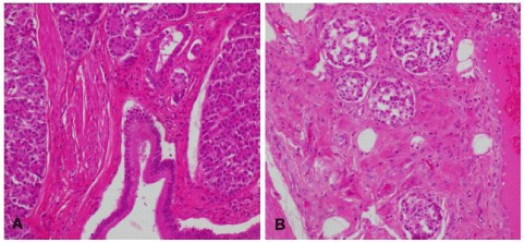
The diagnosis of heterotopic pancreas is difficult as there are no definitive investigative tools. Barium swallow, endoscopic ultrasonography and computed tomography have all been used in aiding diagnosis, but none has proven to be gold-standard [13,14]. Occasionally a diagnosis can be made on the basis of endoscopic biopsies, however, the diagnosis is confirmed only by surgical resection and histopathological examination of the tissue (Figure 4) [2]. The Gaspar Fuentes (1973) classification of heterotopic pancreasis a modified version of the original classification by Heinrich in 1909, dividing the tissue histologically into 4 types. Type 1 consists of acini, ducts and islets similar to the normal pancreas; type 2 consists of ducts only; type 3 consists of acini only; and type 4 consists of islets only [12]. Our case is a Heinrich type 1, which is also the most common type.
A: Exocrine pancreatic tissue. B: Endocrine pancreatic tissue

    Figure 4: Histopathology of the resected tissue.

A: Exocrine pancreatic tissue. B: Endocrine pancreatic tissue
As GISTs are by far the most common gastric submucosal tumour, heterotopic pancreatic tissue can mistakenly be diagnosed as one as it happened in our case. Furthermore, as endoscopic appearance of these heterotopic pancreatic tissues exhibits a well-circumscribed submucosal mass with a normal overlying mucosa, it can further confuse the diagnosis with other submucosal tumours such as GISTs or leiomyomas. However, one feature that distinguishes them apart is that the heterotopic pancreatic tissue may have a central dimpling corresponding with the opening of a duct; although only seen in less than half of cases [12]. Furthermore, pre-pyloric or duodenal location, endoluminal growth pattern, prominent enhancement of overlying mucosa and a CT long diameter to short diameter ratio greater than 1.4 have all been highlighted to favour the characteristics of a heterotopic pancreas over a GIST or leiomyoma [15]. Although this has been proposed, our case had a ratio of 1.3, hence implying that these are not strict criteria.
Most patients with heterotopic pancreas are asymptomatic and require no treatment. However, in patients with severe symptoms, the lesion should be excised either surgically or endoscopically [2]. Due to the difficulty of distinguishing heterotopic pancreatic tissue from primary or metastatic cancer, frozen sections have been recommended to take place routinely during surgical resection to confirm the diagnosis and avoid unwanted radical surgery such as Whipple’s procedure or subtotal gastrectomy [16].
In summary, we have presented an unusual case of a heterotopic pancreatic tissue mimicking a sub-mucosal tumour causing intermittent gastric outlet obstruction. They can be completely asymptomatic or present with abdominal pain, bleeding and obstructive symptoms, and very rarely can undergo malignant transformation. These tissues are a diagnostic dilemma for the clinician and should be considered as a differential diagnosis for GISTs. If symptomatic, surgical resection should be performed.
References
- Christodoulidis, G. Heterotopic pancreas in the stomach: A case report and literature review. World Journal of Gastroenterology. 2007; 13: 6098.
- Shaib YH, Rabaa E, Feddersen RM, Jamal M and Qaseem T. Gastric outlet obstruction secondary to heterotopic pancreas in the antrum: Case report and review. Gastrointestinal Endoscopy. 2001; 54: 527-530.
- Stor Z, Hanzel J. Gastric ectopic pancreas mimicking a gastrointestinal stromal tumour: A case report. International Journal of Surgery Case Reports. 2018; 53: 348-350.
- Filip R, Walczak E, Huk J, Radzki RP, Bienko M. Heterotopic pancreatic tissue in the gastric cardia: A case report and literature review. World Journal of Gastroenterology. 2014; 20: 16779-16781.
- Elpek GO. An unusual cause of cholecystitis: Heterotopic pancreatic tissue in the gallbladder. World Journal of Gastroenterology. 2007; 13: 313.
- Chandan VS, Wang W. Pancreatic Heterotopia in the Gastric Antrum. Archives of Pathology & Laboratory Medicine. 2004; 128: 111-112.
- Dolan RV. The Fate of Heterotopic Pancreatic Tissue: A study of 212 cases. Archives of Surgery. 1974; 109: 762.
- Ormarsson OT, Gudmundsdottir I, Marvik R. Diagnosis and Treatment of Gastric Heterotopic Pancreas. World Journal of Surgery. 2006; 30: 1682-1689.
- Armstrong CP, King PM, Dixon JM & Macleod IB. The clinical significance of heterotopic pancreas in the gastrointestinal tract. The British Journal of Surgery. 1981; 68: 384-387.
- Eisenberger CF, Gocht A, Knoefel WT, Busch CB, Peiper M, Kutup A, et al. Heterotopic pancreas--clinical presentation and pathology with review of the literature. Hepatogastroenterology. 2004; 51: 854-858.
- Zhang Y, Sun X, Gold JS, Sun Q, Lv Y, Li Q, et al. Heterotopic pancreas: A clinicopathological study of 184 cases from a single high-volume medical center in China. Human Pathology. 2016; 55: 135-142.
- Trifan A, Tarcoveanu E, Danciu M, Hutanasu C, Cojocariu C, Stanciu C. Gastric heterotopic pancreas: An unusual case and review of the literature. Journal of Gastrointestinal and Liver Disease. 2012; 21: 209-212.
- Matsushita M, Hajiro K, Okazaki K, Takakuwa H. Gastric aberrant pancreas: EUS analysis in comparison with the histology. Gastrointestinal Endoscopy. 1999; 49: 493-497.
- Cho SJ, Shin KS, Kwon ST. Heterotopic pancreas in the stomach: CT findings. Radiology. 2000; 217: 139-144.
- Kim JY, Lee JM, Kim KW, Park HS, Choi JY, Kim SH, et al. Ectopic Pancreas: CT Findings with Emphasis on Differentiation from Small Gastrointestinal Stromal Tumor and Leiomyoma. Radiology. 2009; 252: 92-100.
- Yuan Z, Chen J, Zheng Q, Huang X, Yang Z, Tang J. Heterotopic pancreas in the gastrointestinal tract. World Journal of Gastroenterology. 2009; 15: 3701-3703.