Case Report
Austin J Surg. 2020; 7(3): 1251.
The Predictability of Laser Assisted Lip Repositioning: A New Evaluation Clinical Scoring Esthetic Scale
Hala H Hazzaa1*, Gasser M Elewa2 and Sherin A Ali3
1Professor of Oral Medicine, Diagnosis and Periodontology, Faculty of Oral and Dental Medicine, Al Azhar University (Girls Branch), Egypt
2Consultant of Laser Applications in Dentistry, General Organization for Teaching Hospitals and Institutes, Egypt
3Associate Professor of Oral Medicine, Diagnosis and Periodontology, Faculty of Dentistry, Tanta University, Egypt
*Corresponding author: Hala H Hazzaa, Professor of Oral Medicine, Diagnosis and Periodontology, Faculty of Oral and Dental Medicine, Al Azhar University (Girls Branch), Cairo, Egypt
Received: July 03, 2020; Accepted: July 28, 2020; Published: August 04, 2020
Abstract
Objective: Lip Repositioning is an invasive surgical method used for correcting the Gummy Smile (GS). Postsurgical relapse is an additional problem that needs to be addressed. Therefore, this bifold6-months study was first conducted to evaluate the Laser-Assisted Lip Repositioning (LALR) as a predictable approach when used in the treatment of patients with hyperactive lip. Additionally, EGD was assessed by a novel Clinical Scoring Esthetic Scale (CSES).
Patients and Methods: This clinical trial was performed on 20 cases with GS using LALR. Esthetic outcomes were assessed using CSES at baseline and 6 months after surgery. This score blindly evaluates the percentages (%) of Excessive Gum Display Difference (EGDD) in each patient. The clinical Evaluation of 5 Lay Persons (ELP) was additionally considered to the display zone of every patient’s spontaneous smile; using a % esthetic scale (0 to 100) by comparing the pre- and post-operative photographs. The EGDD value was added to the % ELP values, then; divided by 2 and the resulting % indicated the clinical grading that was assigned from 1 to 5; with score 1 = Excellent while score 5 = Poor clinical outcome.
Results: The mean scores of CSES of our patients revealed a significant reduction (2.21 ± 1.281) (P-Value ≤ 0.05) at 6-months.
Conclusion: This preliminary clinical report showed predictable results of the laser-assisted lip repositioning technique. The CSES system may be a useful tool to assess the esthetic outcome following lip repositioning procedures.
Keywords: Gummy Smile; Satisfaction; Relapse; Diode Laser
Introduction
Excessive Gingival Display (EGD), commonly termed Gummy Smile (GS), is a condition characterized by an overexposure of the maxillary gingiva while smiling [1,2]. Although the degree of unattractiveness varies between populations, a gingival excess of more than 3 mm is agreed upon worldwide [1-3], with female predominance [4]. GS may result from a single discrepancy, but is more commonly the result of the interplay of multiple factors may be broadly defined as dento-alveolar and nondento-alveolar. Dento-alveolar discrepancies include factors that affect dentition in the form of short clinical crowns, gingival hypertrophy or hyperplasia, altered passive eruption, and extrusion. Nondento-alveolar discrepancies involve Vertical Maxillary Excess (VME) and hyperactive, incompetent, or short lip [5].
Lip Repositioning (LR) procedure was first described in 1973by Rubinstein and Kostianovsky [6] as part of medical plastic surgery. The technique involves a single partial thickness elliptical incision in the depth of the anterior maxillary vestibule and aims to limit smile muscle pull (zygomaticus minor, Levator anguli, orbicularis oris, and levator labii superioris) by reducing the depth of the upper vestibule.
However, the classical LR resulted in increased patient morbidity owing to the scalpel being utilized to remove the strip of the labial mucosa and the frenum. The use of a scalpel resulted in bleeding with decreased visibility in the operatory ?eld, postoperative swelling, and bruising [7], in addition to the postoperative relapse. Several modifications have been therefore introduced to the classical LP approach to improve its clinical outcome, and to minimize its morbidity [8-10]. In this regard, laser-assisted excision serves as an alternate option and provides immediate hemostasis thereby reducing the incidence of hematoma [11]. Although laser assisted approach seems promising in our point of research; literature is sparse related to the clinical application and results obtained [12,13].
Overall, the treatment of GS is mainly indicated for esthetic reasons, to achieve the “Ideal Smile” and patient’s satisfaction. Thus, every effort should be done to give the best chance for our patients to enhance the final esthetic outcomes. However, the esthetic assessment is mainly subjective and might be influenced by cultural background. An objective esthetic evaluation is therefore required to assess the outcomes of plastic surgeries. Taken together, this novel and bifold clinical trial aimed to assess the Laser Assisted Lip Repositioning (LALR) as a predictable method for treating GS-cases; and to additionally propose a Clinical Scoring Esthetic Scale (CSES) as an assessment system for the esthetic outcomes following post-treatment.
Patients and Methods
Study Population
This study was conducted on 20 patients who were treated by LALR during the period from January 2018 to April 2019. Participants were selected from the outpatient clinics of the Oral Medicine and Diagnosis department, Faculty of Oral and Dental Medicine, Al-Azhar University for girls, Egypt; with a common chief complaint of EGD on smiling, with natural anterior dentition and periodontal health. The protocol of the study was approved by the ethical committee of Al-Azhar University for Girls, Egypt.
Extra- and intra-oral examinations were prepared to ensure the suitability of the participants to the study protocol.
Inclusion Criteria
- Participants should be systemically healthy [14].
- Participants had to have at least 3 mm of gingival display on smiling.
- The preoperative lateral cephalometry was assessed to exclude cases with orthognathic problems.
Smokers, pregnant and lactating patients were excluded from this study. In addition, patients with excessive VME were excluded [15].
Preoperative Procedures
Preoperative clinical photographs were taken for each participant. The exact conditions were recorded to ensure reproducibility for postsurgical photographs. The taken photographs included frontal and profile view of relaxed and maximum smile and close-up of lips relaxed and in maximum spontaneous smile.
Measurements of EGD were taken at the maximum smile, and relaxed smile were recorded using a graduated periodontal probe starting from the free gingival margin till the upper lip border.
All participants were asked to fill out preoperative questionnaires to record their own assessment of their smile and later asked to fill out the same questionnaire at the end to record any changes in self-assessment, and evaluate any postoperative complications.
The protocol of the study was explained to all the participating patients, written informed consent was obtained from each case. As part of the routine protocol, oral prophylaxis was done and oral hygiene instructions were given for all the participants. The laser parameters were listed in (Table 1).
Surgical Procedures
- The surgical procedure was initiated after marking the smile line width and height during the patient’s spontaneous smile.
- Adequate infiltration local anesthesia (2% lidocaine with 1:100,000 epinephrine).
- Laser safety protocols were strictly followed. The patient and the clinician were advised to put on the laser safety glasses specific to the wavelength of the laser.
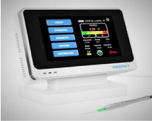
- A 980 nm diode laser (Figure 1) was used for the procedure. 400-µm laser tip in a continuous mode at 0.8 W was first used to demarcate the incision line. The first horizontal incision was outlined at the mucogingival junction and second horizontal incision at about 10 mm parallel to the first incision. Both these outlines were connected at the distal end of the last tooth.
- Laser ablation was carried at an energy setting of 2.17 W average powers in a pulsed mode with pulse duration of 25 ms, using light brush strokes to maintain the depth of ablation, for removal of the epithelial mucosa and exposure of the underlying connective tissue which was followed by removal of tissue tags and saline irrigation.
- The procedure was completed by approximating the midline tissues first, using 4-0 black silk with single interrupted suture (external sutures) to ensure symmetry and proper lip midline placement with the midline of the teeth. The remaining wound margins were approximated with continuous sutures with lock.
    Wavelength
    980 nm


    Power
    6.5    W


    Average    power
    2.17    W


    Fiber    optic tip
    400    microns


    Mode
    Pulsed


    Pulse    duration
    25    ms


    Inter-pulse    duration
    50    ms
Table 1: The used Laser Parameters during in the study.
    Total    number of patients
    Age    (years) Mean± SD


    20
    24.14    ± 1.14


    16/Female
    4/male
SD: Standard Deviation
Table 2: Demographic Data of the study patients.

Figure 1: The Diode Laser Device used in the study.
Postoperative Protocol
Patients were instructed to use:
- Diclofenac potassium, 50 mg, 3 times a day for 1 week.
- Amoxicillin, 500 mg, three times a day for 1 week.
- Chlorhexidine Digluconate mouth wash (0.12%) for 2 weeks.
- Oral hygiene instructions.
Patients were instructed to follow up after 1 week, to assess the wound healing and sutures.
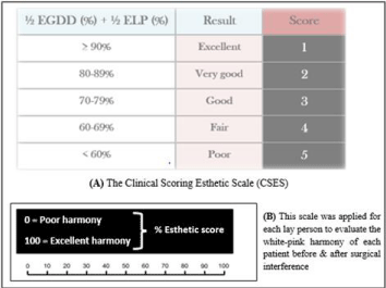
(B) The Evaluation of the Lay Persons (ELP), (n=5) were additionally considered in this scale; this step was based on the harmony between white and pink shadows in the display zone during the spontaneous smile; using a percentage % Esthetic Scale (%ES) after comparing the pre- and post-operative photographs. The sum of (% EGDD + %ES values) was divided by 2 and the resulting % indicated the clinical grading from 1 to 5 according to the figure (score 1: Excellent - score 5: Poor clinical outcome).

    Figure 2: The CSES was set as a blind tool to avoid the biased evaluation after the correction of the gummy smile cases. The percentages (%) of the Excessive Gum Display Difference (EGDD) were calculated for every patient by the same blind investigator who was well trained on the assessment step.
(B) The Evaluation of the Lay Persons (ELP), (n=5) were additionally considered in this scale; this step was based on the harmony between white and pink shadows in the display zone during the spontaneous smile; using a percentage % Esthetic Scale (%ES) after comparing the pre- and post-operative photographs. The sum of (% EGDD + %ES values) was divided by 2 and the resulting % indicated the clinical grading from 1 to 5 according to the figure (score 1: Excellent - score 5: Poor clinical outcome).

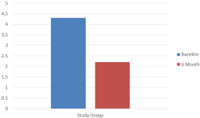
    Figure 3: Bar chart representing change in the mean scores of CSES in the
study group over the follow up intervals.
Clinical Outcomes
Sutures were removed at 2 weeks. Patients were then recalled at 6 months for self-assessment fulfillment, and for taking of photographs, and monitoring of Clinical Scoring Esthetic Scale (CSES); these measures were taken at baseline and 6 months after surgery.
Clinical Scoring Esthetic Scale
The CSES system evaluated 2 main variables at baseline and 6 months following surgery: 1) The EGD in each patient was assessed by a well-trained and non-study periodontist. The percentage (%) of the Excessive Gum Display Difference (EGDD) for each patient was then calculated. 2) The clinical evaluation of the pink and white shadows in the display zone was additionally considered. This parameter was Evaluated by 5 Lay Persons (ELP) who gave their scores using a % Esthetic Scale (%ES) (0-100) by comparing the pre- and post-operative photographs of each case. The sum of the % EGDD value and the % ELP values was divided by 2 and the resulting % indicated the clinical grading that was assigned from 1 to 5; score 1 = Excellent while score 5 = Poor clinical outcome (Figure 2).
Statistical analysis
The collected data was tabulated and analyzed using Statistical Package for Social Science (SPSS 15.0 for windows; SPSS Inc, Chicago, IL, 2001). Data were expressed as mean and Standard Deviation (SD). Paired t-Test was used to compare changes within same group.
For all tests, P ≤ 0.05 was considered significant.
Results
This study included 20 patients, 16females and 4 males aged 21 to 34 years (mean age in years ± standard deviation; 24.14 ± 1.14) (Table 2). They were treated by LALR procedures at the Department of Periodontology, Faculty of Oral and Dental Medicine, Al-Azhar University for girls, Egypt. CSES was used to evaluate the esthetic outcomes of treatment at baseline and 6 months after surgery.
The self-assessment reports reflected the following: All the participants were greatly satisfied of the final outcome in terms of the reduction of EGD at 6 months follow up period. They mentioned no postoperative complications, in terms of infection, pain, bruising and edema; except for only 3 cases who reported postoperative edema for only 1 day (2 patients) 2 days (1 patient).
There was a statistically significant difference within the study group between the baseline and six months follow up assessment of CSES. The mean scores of CSES at baseline and 6-months postoperatively were 4.32 ± 1.08 and 2.21 ± 1.08 respectively; with a significant improvement at P-Value < 0.05 (Figure 3).
Discussion
Currently, the demand for cosmetic procedures has grown exponentially. Excess gingival display is a common esthetic problem that traditionally has been left untreated, unless the associated etiologic factors caused functional challenges. Consistent with a majority of the published clinical studies on LR, our study sample showed a predominantly female population. The predominance of females can be explained by the fact that male patients present lower smile line [16].
The evaluation period used in this study was 6 months from the last surgical interference. Although it seems a relatively short time frame to evaluate the outcomes of LR, 6 months were considered adequate to provide soft tissue stability [2,12].
Lip repositioning was early introduced by Rubinstein and Kostianovsky for the treatment of the EGD cases [6]. Another aggressive approach for treating EGD includes myectomy and partial resection of the levator labii superior is muscle [17]. Lip elevation on smiling can also be limited by placing asilicon spacer between elevator muscles of the lip and the anterior nasal spine [18].
Later, an elliptical-shaped incision at the mucogingival junction and the alveolar mucosa, reflecting a partial thickness flap and removing a strip of the mucosa was proposed for lip repositioning. A combined approach for treating EGD included myotomy of the LLS muscle, subperiosteal dissection of the gingiva, subcutaneous dissection of the lip, and frenectomy [19]. However, a much degree of patient’s morbidity was remarkable.
To overcome these demanding surgical procedures and reduce the morbidity, botulinum toxin was introduced as a minimally invasive alternative to surgical lip repositioning. In this regard, the use of botulinum toxin resulted in satisfactory results due to its ability to block the muscle activity [20, 21]. However, the transitory effect and frequent intramuscular injections forbid the extensive use of botulinum toxin in the correction of EGD.
Most of the aforementioned procedures resulted in complications such as severe discomfort, postoperative bruising, and damage to minor salivary glands [22]. Nevertheless, the major drawback in an esthetic point of view was reports of recurrence of the gummy smile [6]. Taking into consideration these complications, we have used Laser-Assisted Lip repositioning [LALP].
Literature is abundant related to the successful application of diode lasers in soft-tissue surgery; the primary advantages are relatively bloodless surgery with coagulation and reduced bacteremia with minimal discomfort postoperatively. Bleeding associated with classical LR procedures results in hematoma postoperatively, complicating the healing process. This serves as a reservoir for bacteria and tends to loosen the sutures in the initial healing period; the last factor is mentioned as a primary underlying factor for GS relapse. Laser-assisted excision serves as an alternate option and provides immediate hemostasis thereby reducing the incidence of hematoma [23]. Moreover, a bloodless surgical ?eld allows an easy suturing technique which is pivotal to the success of the procedure.
A major bene?t of the laser in our study was relatively less discomfort and edema in the postoperative period. In this regard, laser was compared with the scalpel in another trial; there was reduced intraoperative bleeding and reduced bacteremia which were attributed to sterile in?ammatory condition [24]. An added advantage with the laser was high patient acceptability of the procedure due to its ease and less morbidity and minimal relapse [12], which was in accordance with the results of the patients’ self-assessment reports in this study. Regarding postoperative healing and complications, healing was almost eventful with all participants presenting a singles car line at the mucogingival junction with no postoperative edema; except for three cases only.
The surgical treatment of GS is performed for esthetic reasons: the final outcome should satisfy patients and clinicians. Because the esthetic evaluation is influenced by subjective perceptions [2] by both patients and clinicians, an attempt to minimize the bias of the subjective measures and to categorize objective esthetic assessments can be useful in the evaluation of LR outcomes.
The esthetic outcomes might be influenced by cultural background, and get biased especially if it was completely judged by the patient. Satisfaction assessment is mainly determined by subjective measures using the Visual Analogue Scale (VAS) [1-3]. Although using VAS enables patients to make finely graded assessments, this can have negative effects if questions are unclear or patients feel ambivalent, given that clues on how to formulate an assessment are lacking [25]. A minimum patient ability in terms of visual ability and hand-eye coordination is also required in VAS [26].
In this regard, the authors of this clinical trial introduced a novel clinical esthetic scoring system that assesses the EGD by a well-trained and non-study person, to get reliable and non-biased results [27]. For each patient, the EGD was measured at base line and 6 months by the same investigator; then, the % of EGGD was got for each case.
Another score was taken for each patient by 5 lay persons who were asked to evaluate the patients photographs pre- and post-operatively. The lay persons were non-dental and highly educated [28] who underwent a pre-operative basic knowledge brief course to be oriented by the assessed items and the protocol of the study. Their evaluation was set according to a % ES (0 to 100), to allow easy and wide esthetic perception scale [27]; means were then taken. For each case, the sum of (the % EGDD and the % ELP values) was divided by 2 and the resulting % indicated the clinical grading that was assigned from 1 to 5; score 1 = Excellent while score 5 = Poor clinical outcome.
In this system, the data were documented and the esthetic outcomes were properly evaluated with a negligible bias; it is important that answers are not expressed in an arbitrary manner, but rather that they are assigned to an objective and statistically documentable category. Thus, CSES can be considered a useful alternative to other subjective and self-reported sales for the documentation of the LR outcomes; for future research.
Conclusion
Within the limits of our results, laser-assisted lip repositioning technique is an effective procedure for reducing EGD. A good esthetic outcome was achieved with a remarkable stability at 6 months follow-up. Considering the ease of the procedure, excellent patient acceptability, and providing satisfactory treatment outcome, this can be considered as a feasible alternative in esthetic correction of gummy smile cases. Moreover, the CSES system may be a useful and non-biased tool for assessing the esthetic outcome following lip repositioning procedures.
References
- Panduric DG, Blaskovic M, Brozovic J, Sušic M. Surgical treatment of excessive gingival display using lip repositioning technique and laser gingivectomy as an alternative to orthognathic surgery. J Oral Maxillofac Surg. 2014; 72: 404.
- Tawfik OK, El-Nahass HE, Shipman P, Looney SW, Cutler CW, Brunner M. Lip repositioning for the treatment of excess gingival display: A systematic review. J Esthet Restor Dent. 2018; 30: 101-112.
- Ribeiro-Júnior NV, Campos TV, Rodrigues JG, Martins TM, Silva CO. Treatment of excessive gingival display using a modified lip repositioning technique. Int J Periodontics Restorative Dent. 2013; 33: 309-314.
- Sildeberg N. Goldstein M, Smidt A. Excessive gingival display - Etiology, diagnosis, and treatment modalities. Quintessence Int. 2009; 40: 809-818.
- Robbins JW. Differential diagnosis and treatment of excess gingival display. Pract Periodontics Aesthet Dent. 1999; 11: 265-272.
- Rubinstein A, Kostianovsky A. Cirugiaestetica de la malformacion de la sonrisa. Prensa Med Argent. 1973; 60: 952.
- Ackerman MB, Brensinger C, Landis JR. An evaluation of dynamic lip-tooth characteristics during speech and smile in adolescents. Angle Orthod. 2004; 74: 43-50.
- Miskinyar SA. A new method for correcting a gummy smile. Plast Reconstr Surg. 1983; 72: 397-400.
- Ishida LH, Ishida LC, Ishida J, Grynglas J, Alonso N, Ferreira MC. Myotomy of the levator labii superioris muscle and lip repositioning: A combined approach for the correction of gummy smile. Plast Reconstr Surg. 2010; 126: 1014-1019.
- Ozturan S, Ay E, Sagir S. Case series of laser-assisted treatment of excessive gingival display: an alternative treatment. Photomed Laser Surg. 2014; 32: 517-523.
- Cobb CM. Lasers in periodontics: A review of the literature. J Periodontol. 2006; 77: 545-564.
- Farista S, Yeltiwar R, Kalakonda B, Thakare KS. Laser-assisted lip repositioning surgery: Novel approach to treat gummy smile. J Indian Soc Periodontol. 2017; 21: 164-168.
- Ganesh B, Burnice NKC, Mahendra J, Vijayalakshmi R, Kumar AK. Laser-Assisted Lip Repositioning With Smile Elevator Muscle Containment and Crown Lengthening for Gummy Smile: A Case Report. Clin Adv Period. 2019; 9: 135-141.
- Abramson J. The Cornel Medical index as an epidemiological tool. Am J Public Health Nations Health. 1996; 56: 287-298.
- Alammar AM, Heshmeh OA. Lip repositioning with a myotomy of the elevator muscles for the management of a gummy smile. Dent Med Probl. 2018; 55: 241-246.
- Mazzuco R, Hexsel D. Gummy smile and botulinum toxin: a new approach based on the gingival exposure area. J Am Acad Dermatol. 2010; 63: 1042-1051.
- Miskinyar SA. A new method for correcting a gummy smile. Plast Reconstr Surg. 1983; 72: 397-400.
- Ellenbogen R, Swara N. The improvement of the gummy smile using the implant spacer technique. Ann Plast Surg. 1984; 12: 16-24.
- Ishida LH, Ishida LC, Ishida J, Grynglas J, Alonso N, Ferreira MC. Myotomy of the levator labii superioris muscle and lip repositioning: a combined approach for the correction of gummy smile. Plast Reconstr Surg. 2010; 126: 1014-1019.
- Polo M. Botulinum toxin type A in the treatment of excessive gingival display. Am J Orthod Dentofacial Orthop. 2005; 127: 214-218.
- Suber JS, Dinh TP, Prince MD, Smith PD. On a botulinum toxin A for the treatment of a “gummy smile”. Aesthet Surg J. 2014; 34: 432-437.
- Humayun N, Kolhatkar S, Souiyas J, Bhola M. Mucosal coronally positioned ?ap for the management of excessive gingival display in the presence of hypermobility of the upper lip and vertical maxillary excess: A case report. J Periodontol. 2010; 81: 1858-1863.
- Cobb CM. Lasers in periodontics: A review of the literature. J Periodontol. 2006; 77: 545-564.
- Kalakonda B, Farista S, Koppolu P, Baroudi K, Uppada U, Mishra A, et al. Evaluation of patient perceptions after vestibuloplasty procedure: A Comparison of diode laser and scalpel techniques. J Clin Diagn Res. 2016; 10: ZC96-ZC100.
- Klimek L, Bergmann K, BiedermannT, Bousquet J, Hellings P, Jung K, et al. Visual analogue scales (VAS): Measuring instruments for the documentation of symptoms and therapy monitoring in cases of allergic rhinitis in everyday health care. Allergo J Int. 2017; 26: 16-24.
- Flynn D, van Schaik P, van Wersch A. A comparison of multi-item likert and visual analogue scales for the assessment of transactionally defined coping. Eur J Psychol Assess. 2004; 20: 49-58.
- Hazzaa HH, Attia MS, El Shiekh M, Tawfik MM, El Massieh PMA. A Randomized Placebo-Controlled Intervention with β-Glucan in the Treatment of Localized Aggressive Periodontitis. J Int Acad Periodontol. 2018; 20: 131-142.
- Hass RW, Rivera M, Selvia PG. On the dependability and feasibility of lay persons ratings of divergent thinking. Front Psychol. 2018; 9: 1343-1356.