Clinical Image
Austin J Surg. 2020; 7(3): 1252.
Caseous Annular Calcification of the Mitral Valve
Ermal Likaj*, Selman Dumani, Saimir Kuci and Ali Refatllari
Cardiac Surgery Department, University Hospital Center Mother Theresa, Albania
*Corresponding author: Ermal Likaj, Cardiac Surgery Clinic, Rruga e Dibres, Albania
Received: September 02, 2020; Accepted: September 24, 2020; Published: October 01, 2020
Clinical Image
Caseous Mitral Annular Calcification (CMAC) is a variant of Mitral Annular Calcification (MAC) with a central liquefaction necrosis. The echocardiographic prevalence of CMAC is approximately 0.6% in patients with MAC and 0.06-0.07% in large series of patients of all ages. Optimal treatment of CMAC remains to be established. Although the presence of asymptomatic caseous calcification might be followed-up echocardiographically, any evidence of significant valve dysfunction or embolic phenomena may encourage operative intervention. Uncertain diagnosis may be another indication for surgery.
The importance of correct diagnosis of CMAC extends beyond the possible complications; since several misdiagnoses of CMAC as abscesses and cardiac tumors have been reported leading to inappropriate interventions. Optimal treatment of CMAC remains to be established. We report a case of a 56-year old woman with CMAC diagnosed by trans-thoracic and confirmed by histologic examination after surgery.
The patient complained for chest discomfort and dyspnea for several weeks. Trans-thoracic echocardiogram showed a rounded mass located in the posterior mitral annulus with faint central echo-lucent area. The posterior mitral valve seemed retracted. This prompted the need for trans-esophageal echocardiography which revealed moderate to severe mitral regurgitation and the same characteristics of the mass located in the posterior annulus.

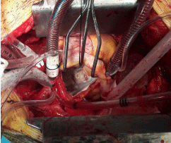
Figure 1: Left atriotomy before the opening of the caseous annular sac.

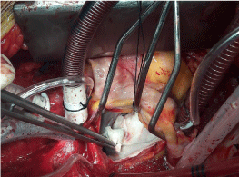
Figure 2: Left atriotomy after the opening of the caseous annular sac.
The patient underwent resection of the mass, closure of the cavity with a pericardial patch and mitral valve replacement. The postoperative period was uneventful. You can observe the interesting images from the left atriotomy before (Figure 1) and after (Figure 2) the opening of the caseous annular sac.
Keywords: Mitral valve; Calcification; Caseous