
Research Article
Austin J Surg. 2021; 8(2): 1268.
Pelvic Congestion Syndrome: Surgical Treatment
Zekilah SR1*, Sallam EM2, Mashaal AB3, Ageez MN4, Dessoky MM5 and Atta IN6
1Department of Vascular Surgery, Alexandria Medical Centre (AMC), Egypt
2Tanta University Hospitals, Lecturer of Vascular Surgery, Egypt
3Department of Gynaecology and Obstetrics, Egypt
4Department of Gynaecology and Obstetrics, Tanta University hospitals, Egypt
5Department of Radiodiagnosis and Ultrasonography, Egypt
6Department of Vascular Surgery, Kafr-Elsheikh University Hospitals, Egypt
*Corresponding author: Samy Rashad Zekilah, Consultant of Vascular Surgery, Alexandria Medical Centre (AMC), Alexandria, Egypt
Received: March 27, 2021; Accepted: April 16, 2021; Published: April 23, 2021
Abstract
Aims: To evaluate the mid and long term efficacy of surgical interruption of the refluxing ovarian veins as a treatment modality for pelvic congestion syndrome.
Study Design: A prospective non comparative interventional study.
Place and Duration of Study: This study was conducted between February 2015 and October 2019 in Alexandria Medical Centre and Tanta Main University Hospital.
Methodology: The study included a 27 patient’s undergone surgical interruption of refluxing ovarian veins with or without sclerotherapy of vulval, perineal or thigh varices, and data were collected prospectively. Detailed history was taken and clinical examination was done for every patient along with routine laboratory investigations and radiological work up was transvaginal and abdominal venous duplex. Follow up was done considering the change in pelvic venous images and pelvic pain scores in comparison to the pre-operative state.
Results: Twenty seven female patients were treated for pelvic congestion syndrome using single session surgical intervention with or without sclerotherapy to pudendal varices. The patients age ranged from 21 to 43 (mean 33.1). All patients presented with chronic continuous pelvic pain. Other associated symptoms as dyspareunia, dysmenorrhea and pudendal varices were found in some cases. Surgical ligation of the ovarian veins were done to all cases, sclerotherapy/ligation of internal iliac varices was done for 6 cases and scerotherapy or surgical interruption of pudendal or thigh varicose veins was done in 21 cases. Technical success was achieved in all patients. Mean pelvic pain score was improved from 7.33 preoperatively to 1.33 and 0.89 in 6 and 12 months of the post-operative recordings. On sonographic basis pelvic reflux disappeared in 26 patients by the end of the follow up. Out of 27 patients treated there were 24 patients satisfied of the procedures at the end of the follow up.
Conclusion: Surgical treatment for pelvic congestion syndrome combined with sclerotherapy to the associated varices was found to be effective, safe and affordable modality of treatment.
Keywords: Pelvic congestion syndrome; Ovarian veins; Pudendal varices; Surgical intervention
Introduction
Chronic pelvic pain can be defined as Pain that may be intermittent or continuous that lasts for 3 months or more, present in the pelvis or pelviabdomenal, usually occurring throughout the menstrual cycle, and not associated with pregnancy [1].
One of the common causes of chronic pelvic pain in women of reproductive age is Pelvic Congestion Syndrome (PCS) which is usually under estimated or misdiagnosed as most of the times pain is attributed to other gynecological disorders, and the patients often suffers from functional disability that interferes with normal life activities and sexual relationship [1].
Venous insufficiency of the Pelvic is assumed to be caused by incompetency of the internal iliac vein, the ovarian vein, or both. It is likely the underlying cause of pelvic congestion syndrome, but the exact etiology is unclear, as It seems to be dependent on multiple factors. The pelvic venous congestion can be caused by hormones, valve insufficiency, or venous obstruction. The release of paininducing substances due to venous congestion along with stasis seems to be the cause of the pain experienced by this patient cohort [2,3].
Notably, about 10% of women in the general population are affected by ovarian varices. Of this 10%, about 60% have PCS [2].
The primary pelvic venous insufficiency is caused by absence of the venous valves or the valvular incompetency. Congenital absence of the ovarian valves was found in 6% of patients on the right side and 13% to 15% on the left side, while valve incompetency was reported in 35% to 46% of patients on the right and 41% to 43% on the left side [4]. The secondary venous incompetency is due to the external compression of the vein like in nutcracker syndrome or May-Thurner syndrome or left renal vein thrombosis (a complication of renal cell carcinoma) [5,6].
The pain manifested with PCS is usually a dull aching pain or a sense of heaviness in the pelvis. It can be unilateral or bilateral or switching from one side to the other. Aggravated with walking, lifting, and longtime standing position. It is often exacerbated before or during the menstruation. Pain intensity worsens with each subsequent pregnancy, also pain usually increases during or after sexual intercourse [7].
Vulvoperineal varicosities can be found in 4 to 8.6% of the patients with ovarian vein insufficiency, thus presenting as an external easily identifiable manifestation of PCS. These varices can extend over the buttock and posteromedial thigh and communicate with both greater and lesser saphenous veins, thus constituting an important potential cause of varicose vein persistence or recurrence after formal saphenous stripping [7].
Patients with PCS, scheduled to undergo intervention, should be investigated to demonstrate the presence of refluxing pelvic veins with ultrasound, retrograde internal iliac or ovarian venography, Computed Tomography (CT), or Magnetic Resonance (MR) imaging [8].
Pelvic duplex ultrasound is considered as the first line imaging study for PCS. As beside giving a detailed anatomy of the ovarian and uterine veins and demonstrating reflux as a retrograde flow in ovarian veins during valsalva, it can exclude other pelvic pathology as a potential cause of chronic pelvic pain like ovarian or uterine masses [9].
Management options include medical treatment (medroxyprogesterone acetate injection, GnRH agonist Goserelin), surgical or laparoscopic ovarian veins ligation or endovascular ovarian vein embolization together with sclerotherapy of vulvoperineal or posteromedial thigh varices if present [3,10-12].
Aim of the work: To evaluate the mid and long term efficacy of surgical interruption of the refluxing ovarian veins as a treatment modality for pelvic congestion syndrome.
Methodology: This is a prospective non comparative interventional study, where a 27 patients undergone surgical interruption of refluxing ovarian veins with or without sclerotherapy of vulval, perineal or thigh varices.
Inclusion criteria: patients presented with manifestations of PCS with or without vulvoperineal or upper thigh varices where pelvic reflux was assumed to be the cause, and duplex study demonstrated the presence of ovarian and broad ligament varices with a diameter ≥ 6mm associated with left or both ovarian veins reflux.
Exclusion criteria:
• Patients with chronic pelvic pain but pathologies other than PCS was discovered as the cause of the pain.
• Patients with pudendal varices but no pelvic symptoms.
• Patients with pelvic varices but ovarian vein reflux cannot be sonographically demonstrated.
• Patients with pelvic varices secondary to common iliac vein obstruction, nut-cracker syndrome, renal tumours or renal vein thrombosis.
• Patient unwilling to do surgery.
Carful history taking was done for each patient regarding the onset and duration of pain. Presence or absence of dyspareunia (painful coitus), dysmenorrhea or external genital varices. The degree of pain and pelvic heaviness were assessed using Pain Score Numerical Rating System (PSNRS) which is a type of visual analogue scores for pain intensity depends on patient questionnaire [13].
Clinical examination included both gynecological and vascular examinations and entails inspection for pudendal or upper thigh varices in the standing position, abdominal palpation and pervaginal palpation where posterior fornix tenderness and pain with mobilization of the cervix were found in most cases.
Pelvi-abdominal venous duplex was our investigation modality using both vaginal and convex abdominal probes to visualize the presence of pelvic varices, refluxing ovarian veins, left renal veins and inferior vena cava and excluding other conditions like ovarian, uterine or renal masses or renal vein thrombosis.
Routine pre-operative laboratory investigations, cardiopulmonary examination, anesthesia consultation were done for each case to prove fitness to undergo surgical intervention.
Surgical intervention: Through standard Pfeninesteil lower abdominal crease incision the abdominal layers were open in order with careful inspection of the outer surface of retro pubic peritoneum as in many cases enlarged varices were found in this location (pudendal and obturator varices) and thought to be the source of vulvoperineal reflux. Division and ligation of these varices was done before opening the peritoneal sac and entering the abdominal cavity. Once the abdominal cavity is entered pelvic structures were carefully examined and any other pathology was identified, identification of the ovaries and ovarian veins running in the suspensory ligament of the ovary. Dissection, Division and ligation with removal of small segments of the varicotic ovarian veins (2 or 3 in number interconnected by small venous channels) with preservation of the ovarian artery, and careful preservation and protection of the ureter which is closely related to ovarian veins in this point. After assurance of hemostasis, closure of the abdomen, no drains were needed (Figures 1-4).
Saphenofemoral disconnection was done via a separate femoral incision for cases proved to have incompetent saphenofemoral junction (s) on either side by preoperative duplex. Sclerotherapy of vulval, pubic or thigh was done at the same cession during the same anesthesia.
Follow up was done with regular post-operative visits at 10 days, one month, 3, 6 and 12 months interval, by means of visual inspection of resolving vulvoperineal varices, the degree of pain resolution was assessed using Pain Score Numerical Rating System [13] (Figure 5), and of course postoperative pelvic duplex.
Results
Twenty-seven patients fulfilled the criteria for enrolment and were included in this study which was conducted between February 2015 and October 2019 in Alexandria Medical Center and Tanta main university hospital, both are in Egypt. Ages of the patients ranged from 21 to 43 years with a mean of 33.1 years (SD: 3.4), all of them were multipara and the number of pregnancies ranged from 2 to 6 with a mean of 3.67 (SD: 0.74).
All patients were suffering from continuous pelvic pain which was moderate to sever in most cases as the pelvic pain score ranged from 6 to 10 with a mean of 9.13 (SD ± 0.32).
On surgical exploration division and ligation of the grossly varicotic ovarian venous plexus which in many cases were thin walled delicate vessels with many interconnecting channels (Figure 1) and in 6 cases, beside ovarian varices, internal iliac varices were also found which were treated by either division and ligation or local injection of AethoxysklerolR foam (to induce spasm of the veins and thus facilitating their dissection without rupture and bleeding) followed by division and ligation with VicrylR sutures (Figure 2-4). Every effort was exerted to identify, protect and avoid injuring or ligation of the ureters which were found to be intimately related to the ovarian venous plexus. Finally dealing with suprapubic, vulval and upper thigh varices with either ligation or sclerotherapy was done, and in 8 cases (28.6%) uni or bilateral saphenofemoral disconnection was done (Figure 6A and 6B) (Table 1).
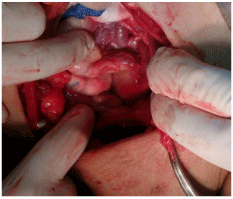
Figure 1: Gross broad ligament varices.
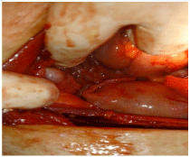
Figure 2: Varicotic internal iliac tributaries.
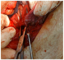
Figure 3: Dissection and separation of ovarian venous plexus.
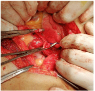
Figure 4: Division of varicotic ovarian veins.
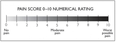
Figure 5: Pain score numerical rating system [13].
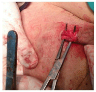
Figure 6A: Dissection of obturator varices causing vulvar varicosities.
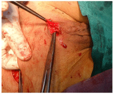
Figure 6B: Dissection of refluxing obturator veins feeding vulvar varices.
![]()
    Procedure done
    Number of cases
    Percentage


    Ovarian    vein (s) division and ligation
        27
    100%


    Internal    iliac varices sclerotherapy/ligation
    6
        22.20%


    Suprapubic    vein ligation
    2
    5.40%


    Vulval/Upper    thigh varices sclerotherapy
    13
    48%


    Saphenofemoral    disconnection with stripping or distal sclerotherapy
    8
    29.60%
Table 1: Procedures done and their percentage.
Follow up visits were scheduled at 10 days (time of stitch removal), one month, 6 months and one year where changes in major preoperative clinical manifestations and duplex criteria were recorded and analysed (Table 2) (Figure 7).
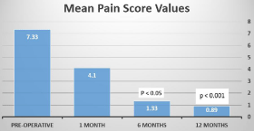
Figure 7: Mean pain score values pre and post-operative.
![]()
    Main manifestation
    Pelvic pain
    Dysmenorrhea
    Dyspareunia
    Pudendal varices
    Reflux on duplex    assessment
    Patients satisfied No of    cases


    Pre-operative
    27
    19
        24
    13
    27
    0


    One    month
    2    (P<0.01)
    3
    -
    6    (sclerosed)
    2    (minor)
        19


    Six    months
        1
    2
    4
    0
        1
        24


    Twelve    months
        1
    2
    4
    0
    1    (P<0.001)
    24    (P<0.05)
Table 2: Main post-operative findings in comparison to pre-operative clinical status.
Discussion
Twenty seven patients may be considered as a small number to be studied in 4 years period but this is due to the strict inclusion criteria for enrollment which necessitate the presence of demonstrable ovarian vein reflux by duplex scanning( trans-abdominal and transvaginal). For the majority of cases pelvic varices was an accidental discovery during investigations of chronic pelvic pain but in others pelvic duplex was done intentionally for suspected pelvic reflux in cases presented with varicose veins in unusual sites. Relatively a considerable number of patients, at the first follow up visit, were still suffering from pelvic pain, dyspareunia and dysmenorrhea and were not satisfied but these parameters improved in subsequent follow up visits. This may be attributed to the recent surgical intervention with violation of the pelvic cavity, besides, pelvic varices seemed to be not completely resolved at this point of time. Painful coitus (dyspareunia) was excluded from the items of follow up in the first visit one month after the operation as the majority of patients avoided any sexual relation in this early post-surgery period for fear of pain but it was present in four patients at the end of the follow up period, and three of them were not satisfied with the whole procedure. This can be explained by either the presence of sexually related psychological problems or the existence of a hidden undiscovered pelvic pathology (for example minor endometriosis or pelvic inflammatory disease). Almost all recent series, published in English literature, adopted the trans catheter embolization of ovarian veins or internal iliac vein tributaries in managing patients suffering from PCS caused by pelvic varices, thus investigators in these series could not perform direct exploration and visualization of the pelvic contents for detecting or excluding any concomitant pathology (apart from few case series in which laparoscopic exploration with ovarian veins clipping was done). The lower cost, minimal morbidity with almost no mortality and availability in most centers may make surgery an appealing option. Both ligation and sclerotherapy could be done during the surgical intervention for controlling thin walled fragile vessels and both of them are cost effective, furthermore, any associated pudendal or lower limb varices can be treated by either surgery or sclerotherapy during the same anesthesia. The excellent cosmetic result of the Pfeninesteil lower abdominal crease incision together with the existence of previously old cesarian section scares in many cases makes the cosmetic issue not a point of concern. Series describing surgical management of pelvic varices are very scanty in the literature, but one report by Gargiulo et al. [14] in which 23 patients were treated by laparoscopic high ovarian vein(s) clipping and achieved a 60-90 % resolution of pelvic varices over 12 months.in the current study low ligation of ovarian veins (in the suspensory ligament of the ovary) was done which seemed adequate via the cosmetic lower abdominal crease incision, besides, internal iliac and obturator varices could be dealt with through the same approach. Drazic et al. [15] and Cordts et al. [16] reported for the coil embolizaion of ovarian veins for 17 and 11 patients respectively. Duplex assessment was the primary diagnostic modality for Drazic and his group and they achieved a 100% symptomatic relieve over 32 months of follow up after ovarian vein embolization while for Cordts and his group symptomatic relieve of 40-100 % was noted with ovarian and obturator veins embolization at a mean follow up period of 13.4 months. Results of both studies were comparable to the results of the current study but with longer follow up periods.
Conclusion: Surgical treatment for pelvic congestion syndrome combined with sclerotherapy to the associated pudendal and upper thigh varices was found to be effective, safe and affordable modalities of treatment with low morbidity and almost no procedure related mortalities. Its fair results, cost effectiveness and low recurrence rate may make more popular in the future especially in developing countries.
References
- Koo S, Fan CM. Pelvic congestion syndrome and pelvic varicosities. Tech Vasc Interv Radiol. 2014; 17: 90-95.
- Daniels J, Gray R, Hills RK, Latthe P, Buckley L, Gupta J, et al., LUNA Trial Collaboration. Laparoscopic uterosacral nerve ablation for alleviating chronic pelvic pain: a randomized controlled trial. JAMA. 2009; 302: 955-961.
- Soysal ME, Soysal S, Vicdan K, Ozer S. A randomized controlled trial of goserelin and medroxyprogesterone acetate in the treatment of pelvic congestion. Hum Reprod. 2001; 16: 931-939.
- Ahlberg NE, Bartley O, Chidekel N. Right and left gonadal veins. An anatomical and statistical study. Acta Radiol Diagn (Stockh). 1966; 4: 593- 601.
- Birn J, Vedantham S. May-Thurner syndrome and other obstructive iliac vein lesions: meaning, myth, and mystery. Vasc Med. 2015; 20: 74-83.
- Gulleroglu K, Gulleroglu B, Baskin E. Nutcracker syndrome. World J Nephrol. 2014; 3: 277-281.
- Jung SC, Lee W, Chung JW, et al. Unusual causes of varicose veins in the lower extremities: CT venographic and Doppler US findings. Radiographics. 2009; 29: 525-536.
- Gloviczki P, Comerota AJ, Dalsing MC, Eklof BG, Gillespie DL. The care of patients with varicose veins and associated chronic venous diseases: clinical practice guidelines of the Society for Vascular Surgery and the American Venous Forum. J Vasc Surg. 201; 53: 2S-48S.
- Leung SW, Leung PL, Yuen PM, Rogers MS. Isolated vulval varicosity in the non-pregnant state: a case report with review of the treatment options. Aust NZJ Obstet Gynaecol. 2005; 45: 254-256.
- Farquhar CM, Rogers V, Franks S, Pearce S, Wadsworth J, Beard RW. A randomized controlled trial of medroxyprogesterone acetate and psychotherapy for the treatment of pelvic congestion. Br J Obstet Gynaecol. 1989; 96: 1153-1162.
- Gandini R, Konda D, Abrignani S, Chiocchi M, Da Ros V, Morosetti D, et al. Treatment of symptomatic high-flow female varicoceles with stop-flow foam sclerotherapy. Cardiovasc Intervent Radiol. 2014; 37: 1259-1267.
- Bittles MA, Hoffer EK. Gonadal vein embolization: treatment of varicocele and pelvic congestion syndrome. Semin Intervent Radiol. 2008; 25: 261-270.
- Williamson A. Pain: a review of three commonly used pain rating scales. Issues in Clinical Nursing. 2005; 14: 798-804.
- Gargiulo T, Mais V, Brokaj L. Bilateral laparoscopic transperitoneal ligation of the ovarian veins for treatment of pelvic congestion syndrome. J Am Assoc Gynacol Laparosc. 2003; 10: 501-504.
- Drazic BO, Zárate BC, Valdés EF, Mertens MR, Bergoeing RM, Krämer SA, et al. Embolization of insufficient pelvic veins for pelvic congestion syndrome. Analysis of 17 cases. Rev Med Chil. 2019; 147: 41-46.
- Cordts PR, Eclavea A, Buckley PJ, DeMaioribus CA, Cockerill ML, Yeager TD. Pelvic congestion syndrome: early clinical results after transcatheter ovarian vein embolization. J Vasc Surg. 1998; 28: 862-868.