Case Report
Austin J Surg. 2022; 9(1): 1285.
Biliary Cystadenocarcinoma Misdiagnosed as a Hydatic Cyst of the Liver: About a Case and Literature Review
Youssef El Mahdaouy*, Noureddine Njoumi, Mohammed El Fahssi, Mbarek Yaka, Abderrahman Hjouji and Abdelmounaim Ait Ali
Department of Surgery, Visceral Surgery Service 2, Mohamed V Military and Training Hospital, Rabat, Morocco
*Corresponding author: Youssef El Mahdaouy, Department of Surgery, Visceral Surgery Service 2, Military and Instruction Hospital Mohamed V, Rabat, Morocco
Received: May 19, 2022; Accepted: June 21, 2022; Published: June 28, 2022
Abstract
Biliary cystadenocarcinomas are very rare malignant epithelial tumors of the liver. The preoperative diagnosis of this cystic tumor is difficult because its clinical and radiological presentation is non-specific.
Complete surgical removal is the recommended treatment for hepatic cystadenocarcinoma. Incomplete resection is a source of tumor recurrence and metastatic dissemination, which is the case when it is wrongly diagnosed as another benign cystic lesion of the liver.
We report a new case of biliary cystadenocarcinoma that has been diagnosed as a hepatic hydatic cyst, resulting in inadequate management.
Keywords: Biliary Cystadenocarcinoma; Hepatic Hydatic Cyst; Cystic Lesion of the liver
Introduction
Biliary cystadenocarcinomas are very rare epithelial malignant liver tumors, only 247 cases have been reported in the literature [1].
The absence of clinical, biological and radiological specificity makes pre-operative and even intraoperative diagnosis difficult. Thus, biliary cystadenocarcinoma may be taken for another cystic liver lesion.
The definitive diagnosis is made in the pathological study of the lesion after complete surgical resection, the only potentially curative treatment.
The rarity and diagnostic difficulty of this pathology prompted us to report a new case of liver cystadenocarcinoma initially wrongly diagnosed as a hepatic hydatic cyst, especially since the hydatic cyst of the liver is a cystic lesion common epidemiologically in Morocco.
Case Report
This is a 67-year-old patient, housewife, with a history of iterative hepatic surgery for hydatic cyst with negative serology: salient dome resection in 2013 and 2015, and left hepatic resection (left lobectomy?) for a new hydatic recurrence in 2018 whose histopathological analysis returned in favor of a liver cystadenoma in high-grade dysplasia associated with the presence of two microfoci of an infiltrating adenocarcinoma.
In 2019, the patient consults for abdominal pain in the right hypochondria without other associated signs, hence her admission to the service for specialized management.
The general examination finds a patient in general condition preserved, hemodynamic and respiratory stable, apyretic and her conjunctiva are normally colored. The physical examination reveals, besides the scar of the previous operations under the right rib, a soft abdomen to palpation.
The standard bioassay: complete blood count, coagulation test, blood ionogram, renal function and liver test have returned to normal. Serum tumour marker levels (CA19-9, CA 125, ACE and AFP) are normal.
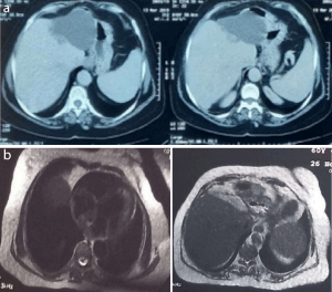
Abdominal computed tomography (CT) was performed and resulted in two hepatic cystic masses in the left liver, of tissue density, homogeneous content and well limited wall, without vegetations or partitions, successively measuring 07 04 and 04 04 04 cm enhanced after injection of contrast product (Image a). Hepatic magnetic resonance imaging (MRI) found a cystic lesion with a long axis of 07cm, this lesion is contiguous to another lesion with a long axis of 04 cm, which has an enhanced irregular thick wall with diffusion restriction (Image b).

Image a & b: CT and MRI results showing tumor recurrence.
The diagnosis of a recurrent cystic tumor of the liver, in the case of biliary cystadenocarcinoma, was therefore selected. tumor is limited to the live at staging.
The surgical exploration noted the presence of a cystic mass in the old liver cross section, adhering to the small gastric curvature, the dissection of which required resection of the gastric serosa. A left hepatectomy was performed by taking the tumor masses in monobloc. The surgical follow-ups were simple.
Six months later, the general condition of the patient worsened with recurrence of pain in the right hypochondria and installation of jaundice related to the progression of the disease objectified to imaging (CT). The patient died four months after the start of chemotherapy.
Discussion
Biliary cystadenocarcinoma is one of the malignant epithelial tumours of the liver. It is a very rare tumour with an incidence of less than 0.41% of malignant epithelial tumours [2,3].
Unlike biliary cystadenoma, which occurs almost exclusively in women of average age of 45 years, biliary cystadenocarcinoma occurs in both sexes with an almost equal prevalence and at a higher mean age of 55 years [1].
Biliary cystadenocarcinoma most likely develops from focal or diffuse malignant degeneration of pre-existing biliary cystadenoma with mesenchymal ovarian-type stroma.3, 4 20-25% of these tumors degenerate into biliary cystadenocarcinoma according to the natural history of all adenocarcinomas with progression from benign epithelium to carcinoma through atypia and dysplasia [1,3].
Rarely, biliary cystadenocarcinoma can develop from ectopic remains foregut sequestered in the liver, congenital liver cyst, common bile duct and gallbladder [3,4].
The wide etiological range of hepatic cystic lesions and the lack of specificity of the clinical, biological and radiological presentation of biliary cystadenocarcinoma make it difficult to diagnose preoperatively [1,3].
Indeed, the symptoms of biliary cystadenocarcinoma are inconstant and non-specific. It is a tumor of slow evolution, it can remain asymptomatic and in this case of incidental discovery at the imaging requested for other pathologies or even at autopsy. Symptomatic patients usually complain of abdominal pain in the right hypochondria, abdominal distension or palpable mass. Jaundice, fever, ascites and weight loss are less frequent and generally late symptoms related to a progression of the disease (tumor recurrences or distal metastases). Complications have been reported, including bleeding, ruptures and cystic infections [3].
Biologically, haematological and biochemical tests are usually normal. Sometimes cholesatase is noted. Tumor markers are not specific. Increased serum levels of these markers were also found in biliary cystadenomas. In contrast, high serum levels of ACE and CA 19-9 have been reported to be more suggestive of biliary cystadenocarcinoma. Finally, the diagnosis of biliary cystadenocarcinoma cannot be ruled out if the serum level of these markers is normal [1].
Preoperative diagnosis of biliary cystadenocarcinoma is mainly based on imaging results [1,5]. Its discovery is easily made by ultrasound. It demonstrates the cystic character of the lesion, which is often voluminous and thick-walled. The lesion can be single or multilocular with irregular septations, papillary projections and mural nodules. Parietal or septal calcifications can be seen but are rare and would be a diagnostic element in favor of biliary cystadenocarcinoma [1,2]. Contrast-enhanced ultrasound can be informative with regard to the vascularization of solid parts of a cystic lesion [6].
Abdominal CT, performed without contrast injection, confirms the cystic nature of the lesion. Typically, it is a solitary multilocular or unilocular lesion with well-circumscribed smooth edges and internal septations. The contrast product injection shows an enhancement of the wall, septas and mural nodules highlighting them better. Calcifications can be observed within the wall and septas in a minority of cases [1,2].
Hepatic MRI, in addition to ultrasound and CT, shows a multilocular and well limited cystic mass with enhancement after intravenous injection of Gadolinium. It also allows a characterization of the intracystic content with variation of the signal level in the different loculi on the T1 and T2-weighted sequences [3,5].
Although the presence of complex and especially hemorrhagic fluid content, mural nodules and calcifications along the wall or septas strongly support the diagnosis of biliary cystadenorcinoma, its differentiation from biliary cystadenoma often remains difficult due to the large overlap of radiological characters [1,5].
Ultrasound percutaneous biopsy is not recommended because it rarely allows a definitive diagnosis and carries a risk of peritoneal dissemination in case of malignancy. Ultrasound percutaneous aspiration puncture of cystic contents is of no interest; in addition to the risk of dissemination, no malignant cells were recovered in patients with biliary cystadenocarcinoma who underwent a peroperative cytological examination. Studies have also shown that there is no statistically significant difference in tumour marker levels in the blood or cystic fluid [1].
Despite the progress and performance of current imaging means, preoperative diagnosis of biliary cystadenocarcinoma remains difficult (low specificity) [1].
Thus, given the risk of recurrence and especially the metastatic dissemination, the differential diagnosis of biliary cystadenocarcinoma of other hepatic cystic lesions is a very important step. It is done mainly with biliary cystadenoma, hydatic cyst, complicated biliary cyst, alveolar echinococcosis, hepatic abscess, hematoma remodeled and less frequently with necrotic metastases [5].
Hydatic cyst of the liver is a major public health problem in endemic countries such as the Mediterranean and North African countries such as Morocco. It is secondary to accidental contamination by echinoccocus granulosus. Hepatic hydatic cyst may share the same clinical presentation with biliary cystadenocarcinoma. Hydatic serology can be negative in 10% of cases and biliary cystadenocarcinoma may be misdiagnosed as a hydatic cysts of liver type III (cystic lesion containing daughter vesicles) and especially hydatic cysts of liver type IV (heterogeneous lesion with irregular wall) Gharbi [5]. The patient’s hepatic cystic lesion was incorrectly diagnosed as a hydatic cyst of the liver.
Even in the operating room, the diagnosis can remain problematic; usually not evoked goes the rarity of this pathology or taken for another cystic lesion. Thus the surgeon, by adopting an easy surgical attitude due to lack of expertise in hepatobiliary surgery can worsen the prognosis of patients with hepatic cystadenocarcinoma by inadequate treatment [1,7].
The radical excision by a complete formal hepatic resection with a margin of normal liver tissue (1 cm) is the only potentially curative treatment when hepatic cystadenocarcinoma is suspected [3]. The enucleation of the cyst, as performed for biliary cystadenoma, is not recommended as there is an increased risk of recurrence. Authors discuss this technique even in cases of biliary cystadenoma [1]. There is no indication of adjuvant treatment if the tumor is limited to the liver. Chemotherapy and/or radiation therapy may be indicated in case of metastases (20%) [3].
With a recurrence rate of 4.8% and a mortality rate of 24.2%, the prognosis of biliary cystadenocarcinoma is better than other malignant epithelial tumours of the liver [1]. the survival rate reported in the literature varies from 25% a100% to 5 years.4 Also it has been reported that the absence of the mesenchymal stroma (ovarian type) in biliary cystadenocarcinoma is associated with a poor prognosis especially in men and even in cases of complete resection [4].
Conclusion
Given its rarity and the lack of specificity of its clinical, biological and radiological presentation, in addition to the wide etiological range of liver cystic masses, in our contests the hydatic cyst of the liver, Preoperative diagnosis of hepatic cystadenocarcinoma is a real challenge.
We propose a radical excision of all multilocular cystic tumours of the liver.
References
- Klompenhouwer AJ, Cate DWGT, Willemssen FEJA, Bramer WM, Doukas M, Man RAD, et al. The impact of imaging on the surgical management of biliary cystadenomas and cystadenocarcinomas; a systematic review. HPB : the official journal of the International Hepato Pancreato Biliary Association. 2019; 21(10): 1257-1267. doi:10.1016/j.hpb.2019.04.004.
- M. Souei Mhiri, K. Graiess Tlili, M.T. Yacoubi, À propos d’un cas de cystadénocarcinome biliaire, Journal de Radiologie, 2005; 86(9): 1035-1037. https://doi.org/10.1016/S0221-0363(05)81488-X.
- Läuffer JM, Baer HU, Maurer CA, Stoupis C, Zimmerman A, Büchler MW. Biliary cystadenocarcinoma of the liver: the need for complete resection. European journal of cancer. 1998; 34(12): 1845-1851. doi:10.1016/S0959- 8049(98)00166-X.
- Vogt DP, Henderson JM, Chmielewski E. Cystadenoma and cystadenocarcinoma of the liver: a single center experience. Journal of the American College of Surgeons. 2005; 200(5): 727-733. doi:10.1016/J. JAMCOLLSURG.2005.01.005.
- Labib PLZ, Aroori S, Bowles M, Stell D, Briggs C. Differentiating Simple Hepatic Cysts from Mucinous Cystic Neoplasms: Radiological Features, Cyst Fluid Tumour Marker Analysis and Multidisciplinary Team Outcomes. Digestive Surgery. 2016; 34(1): 36-42. doi:10.1159/000447308.
- Lin M, Xu H, Lu M, Xie X, Chen L, Xu Z, et al. Diagnostic performance of contrast-enhanced ultrasound for complex cystic focal liver lesions: blinded reader study. European Radiology. 2008; 19(2): 358-369. doi:10.1007/ s00330-008-1166-8.
- Ahmad Z, Uddin N, Memon W, Abdul-Ghafar J, Ahmed A. Intrahepatic biliary cystadenoma mimicking hydatid cyst of liver: a clinicopathologic study of six cases. Journal of Medical Case Reports. 2017; 11(1). doi:10.1186/s13256- 017-1481-2.