
Case Report
Austin J Surg. 2022; 9(2): 1287.
A Case Report: Massive Spontaneous Hemorrhage of Neurofibromatosis Type 1 on Upper Back
Yeh CC*
Division of Plastic Surgery, Department of Surgery, Chi-Mei Medical Center, Taiwan
*Corresponding author: Chin-Choon Yeh, Division of Plastic Surgery, Department of Surgery, Chi-Mei Medical Center, Yong-Kang Distinct, Tainan City, Taiwan
Received: July 07, 2022; Accepted: August 05, 2022; Published: August 12, 2022
Abstract
Neurofibromatosis type 1 (NF1) (von Recklinghausen’s disease) is an autosomal dominant disorder characterized by café-au-lait spots, pigmented hamartomas of the iris, and multiple neurofibromas. Patients can present with hemorrhage secondary to trauma or rarely with spontaneous hemorrhage.
We report a 42-year-old man with long-term history of a giant neurofibroma on his back who suffered from spontaneous hemorrhage. Series examinations and treatments were given to stop bleeding and eventually to heal the wound.
Keywords: Neurofibromatosis type 1; Spontaneous hemorrhage
Introduction
Neurofibromatosis ype 1 (NF1), also called as von Recklinghausen’s disease, usually involves organs of ectodermal oringin. It is an autosomal dominant disease having an incidence of 1 in 3000 individuals. It primarily involves the peripheral nervous system and usually presents with many neurofibromas [1]. Cutaneous neurofibromas are hallmarks of Neurofibromatosis type 1 (NF1) and cause the main disease burden in adults with NF1 [2]. People suffering from neurofibromatosis face various challenges in their daily activities, including spontaneous rupture and hemorrhage especially on large-sized lesions.
Case Report
A 42-year-old man has history of neurofibromatosis type 1 for more than 30 years was admitted to the hospital due to an emergent episode of massive spontaneous hemorrhage inside a giant neurofibroma on the left side upper back. The patient had a left upper back neurofibroma with size about 6 cm for long time, and then it got enlargement to 14x8-cm within 6 hours. There were no traumas or fierce exercise before the episode. On physical examination, it revealed that patient was of short stature with severe scoliosis, and displayed diffuse cafe-au-lait spot on the skin, plexiform neurofibroma on the back. There was a 14x8-cm hematoma originating from neurofibroma on the left side back (Figure 1). Local tenderness and 5x3 cm skin erosion on neurofibroma were also found (Figure 2). Tachycardia with acceptable blood pressure were recorded. Trunk CT showed that at left scapula area, a hematoma with considering active bleeding from left thoracodorsal artery was favored (Figure 3). At emergency room, hematoma compression with bandage, fluid resuscitation and pain control with analgesics were managed. 4 hours later however, dizziness, unstable systolic blood pressure, increasing back hematoma size (from 14x8-cm to 20 x 18- cm), hemoglobin level progressive decrease (from 137 g/L to 84 g/L) occurred. Urgent angiographic embolization was performed to stop the bleeding. During left subclavian arteriography, it proved that the large hematoma was truly supplying by branch of left thoracodorsal artery. Major feeder was embolized with gel-foam pieces and a 0.018/14cm/4mm coil (Figure 4). The patient’s hemodynamic condition stabilized gradually. 4 days after admission, severe sepsis due to neurofibroma wound skin necrosis causing soft tissue infection was noted (Figure 5). Clinical condition subsided after systemic antibiotics administration. 11 days after admission, the hematoma and neurofibroma were surgically resected, with 1800 gm hematoma and some active bleeder from latissimus dorsi muscle seen during operation (Figure 6). In spite upper back sutured wound had partial skin necrosis post-operatively, regular wound care with Neomycin containing ointment was applied. The wound healed day by day. The patient was discharged 20 days after the operation. There was no recurrence of neurofibroma or hematoma at 3-month follow-up (Figure 7).
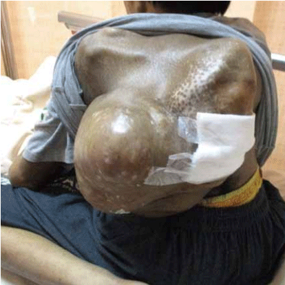
    Figure 1: There was a 14x8-cm hematoma originating from neurofibroma on

the left side back.
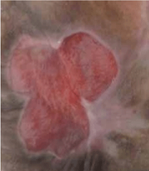
    Figure 2: Local tenderness and 5x3 cm skin erosion on neurofibroma were

also found.
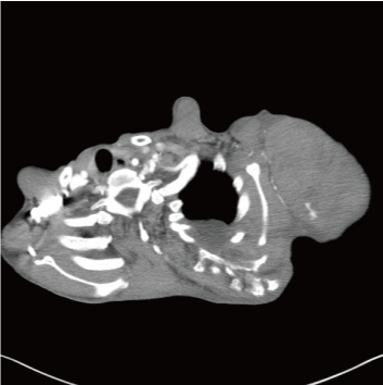
Figure 3: Active bleeding from left thoracodorsal artery.
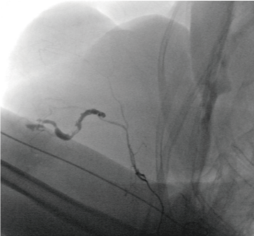
    Figure 4: Major feeder was embolized with gel-foam pieces and a

0.018/14cm/4mm coil.
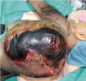
Figure 5: Skin necrosis causing soft tissue infection.
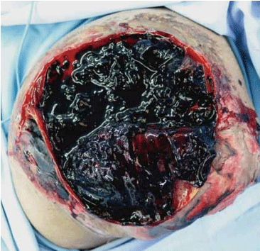
Figure 6: Hematoma and some active bleeder from latissimus dorsi muscle.
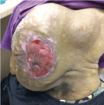
Figure 7: 3-month follow-up.
Discussion
Neurofibromatosis type 1 (NF1) and Neurofibromatosis type 2 (NF2) are autosomal dominant inherited neurocutaneous disorders or phakomatoses secondary to mutations in the NF1 and NF2 tumor suppressor genes, respectively. Although they share a common name, NF1 and NF2 are distinct disorders with a wide range of multisystem manifestations that include benign and malignant tumors. Imaging plays an essential role in diagnosis, surveillance, and management of individuals with NF1 and NF2. Therefore, it is crucial for radiologists to be familiar with the imaging features of NF1 and NF2 to allow prompt diagnosis and appropriate management. Key manifestations of NF1 include cafe-au-lait macules, axillary or inguinal freckling, neurofibromas or plexiform neurofibromas, optic pathway gliomas, Lisch nodules, and osseous lesions such as sphenoid dysplasia, all of which are considered diagnostic features of NF1. Other manifestations include focal areas of signal intensity in the brain, low-grade gliomas, interstitial lung disease, various abdominopelvic neoplasms, scoliosis, and vascular dysplasia [3].
Multidimensional imaging’s using 3/2D ultrasound, CTA, and DSA plays a crucial role in assessing the source of bleed and planning intervention. Neuroimaging findings in patients with NF1 include focal areas of increased signal intensity on T-2 weighted imaging , called ‘unidentified bright objects’ and may disappear in adulthood. In 2–6% of patients with NF1, increased brain volume and abnormal cerebral vessels are also detected [4].
Definite treatment to prevent re-bleeding is considered to be surgical operation including total/subtotal resection and vessel ligation in literatures [1,5,6].
Conclusion
As to our knowledge, massive spontaneous hemorrhage in the soft tissue of upper back is very rare in patients with NF1. This case shows that combination of angiographic embolization and surgical excisions have great effectiveness in control of massive hemorrhage for patients with NF1.
References
- Aljindan F, Aljehani L, Alsharif B, Mortada H. Management of Neurofibromatosis of the Nipple-Areolar Complex. Case Reports in Surgery. 2021; 2021: 1-4.
- Kallionpää RA, Ahramo K, Martikkala E, Fazeli E, Haapaniemi P, Rokka A, et al. Mast Cells in Human Cutaneous Neurofibromas: Density, Subtypes, and Association with Clinical Features in Neurofibromatosis. Dermatology. 2021; 238: 329-339.
- Mindy X Wang, Jonathan R Dillman, Jeffrey Guccione, Ahmed Habiba, Marwa Maher, et al. Neurofibromatosis from Head to Toe: What the Radiologist Needs to Know. Radiographics. 2022; 42: 1123-1144.
- Edwin Stephen, Rajeev Kariyattil, Alok Mittal, Faisal Al-Azri, Khalifa Al- Wahaibi, et al. Spontaneous Near Fatal Hemorrhage into Neurofibromatosis Type 1 Lesion in the Scalp. Oman Med J. 2022; 37: e387.
- Solares, I., D. Vinal, and M. Morales-Conejo, Diagnostic and follow-up protocol for adult patients with neurofibromatosis type 1 in a Spanish reference unit. Rev Clin Esp (Barc). 2022.
- Sone M, Takenaka Y, Miyabe C, Suzuki M, Nakao M, Hagiwara N, et al. Ischaemic skin ulcers with neurofibromatosis 1 successfully treated with stent implantation. Clinical and Experimental Dermatology. 2021; 47: 459-461.