Case Report
Austin J Surg. 2022; 9(3): 1292.
A Case of Retroperitoneal Panniculitis with Paralytic Ileus Mimiking a High Intestinal Obstruction
Rahali A*, Njoumi N, Elfahssi M, Elhjouji A, Zentar A and Ait Ali A
Department of Visceral Surgery, Mohammed V Military Teaching Hospital, Rabat, Morocco
*Corresponding author: Anwar Rahali, Department of Visceral Surgery II, Mohammed V Military Teaching Hospital, Rabat, Morocco
Received: October 10, 2022; Accepted: November 04, 2022; Published: November 11, 2022
Abstract
Retroperitoneal panniculitis is a rare, benign and nonspecific inflammatory disease that affects the retroperitoneal adipose tissue. The specific cause of the disease is unknown. The diagnosis is evoked by computed tomography and is rarely confirmed by biopsies. Treatment is based on a few selected immunosuppressive drugs. Surgical resection is sometimes attempted for complicated forms, although the surgical approach is often limited. We report a case of a 22-year-old man diagnosed with retroperitoneal panniculitis complicated by a paralytic ileus and mimicking an acute abdomen. A self-limiting course of disease was obtained by adopting a conservative approach.
Introduction
Retroperitoneal panniculitis is a rare benign disease of unknown etiology, characterized by non-specific inflammation of retroperitoneal adipose tissue. The pathophysiology of this lesion remains unclear, however, few reports suggest hypothesis which may be useful in the early diagnosis and for elucidating the pathophysiology [1]. CT features of the retroperitoneal panniculitis, usually highly suggestive, have recently been described clearly. CTguided or laparoscopic biopsies seem rarely necessary to confirm the diagnosis [1-2].
We present details of a case of a 22-year-old man diagnosed with retroperitoneal panniculitis as well as a literature review to compare various presentations, etiologies and potential treatment modalities.
Case Report
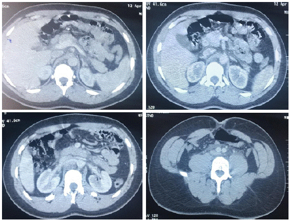
A 22-year-old man developed acute onset of perpetual epigastric pain and intermittent episodes of postprandial vomiting and nausea, and presented to the emergency department 24 h after the beginning of symptoms. There was no significant medical or family history. A physical examination revealed marked epigastric tenderness accompanied by abdominal distension without muscular defense. A diagnosis of acute pancreatitis was suspected. However, the blood examination revealed a normal lipase and amylase levels. An abdominal contrast-enhanced CT showed a high-density lesion of retroperitoneal panniculus adiposus tissue especially behind the pancreas, with a paralytic ileus (Figure 1).

Figure 1: Transverse CT scan of the abdomen showing a high-density lesion within the retroperitoneal space.
A final diagnosis of retroperitoneal panniculitis was considered. The symptoms gradually disappeared and white cell count and C-reactive protein returned to normal on day 5. The patient was discharged on day 8 after receiving conservative treatment. After 20 days, there were no abnormal findings on CT and the patient was referred to internal medicine consultation for additional care.
Discussion
Retroperitoneal panniculitis is a rare benign disease characterized bychronic and nonspecific inflammation of retroperitoneal adiposus tissue. Abdominal panniculitis was first reported by Jura in 1924 in mesenteric fat tissue. However, few cases with retroperitoneal panniculitis have been previously described. It usually affects the men with a male/female ratio of 1.8 and its incidence increases with age [1-3].
The causes and pathogenesis of retroperitoneal panniculitis are unknown. Infection, autoimmune disease, malignant neoplasm and previous abdominal surgery have been implicated by some authors [4]. Our patient was considered to have an idiopathic etiology by reason of the self-limited course.
The disease is often asymptomatic. When present as in the case of our patient, clinical symptoms are non-specific and include abdominal pain, abdominal fullness, nausea, vomiting, anorexia, pyrexia, change in bowel habit and weight loss [5]. Physical examination may contain a palpable mass, abdominal tenderness, abdominal distension, muscular defense and as cites [5-6].
Laboratory profile of retroperitoneal panniculitis is nonspecific and generally unhelpful [6-7]. However, with the arrival of latest imaging technology like high-resolution CT or magnetic resonance imaging, distinguishing retroperitoneal panniculitis from other retroperitoneallesions with similar imaging features such aslymphoma, lymphosarcoma, liposarcoma, desmoid, and metastatic neoplasm seems feasible [8-9]. The imaging appearance of retroperitoneal panniculitis is visualized usually as a high-density heterogeneous lesion of retroperitoneal panniculus adiposus tissue. Calcification or a fibrous capsule may be seen and generally no invasion of adjacent structures is present [5-10].
Histological examination is sometimes recommended to confirm the diagnosis especially for symptomatic and chronic cases. Multiple CT-guided or laparoscopic biopsies are requiredas an alternative to laparotomy [11].
Retroperitoneal panniculitis resolves spontaneously in the majority of cases and it often runs a self-limiting course. Recurrences are very exceptional. Our patient developed a paralytic ileus mimicking a high intestinal obstruction. Other rare complications are reported such as ureteral obstruction, reactive pancreatitis and ascites [1-11-12].
Treatment is individualized on a case by case basis. The limited proportion of cases with persistent symptoms responds well to immunosuppressive therapy (corticosteroids, azathioprine, thalidomide, colchicine or cyclophosphamide). As a form of therapy for retroperitoneal panniculitis, surgery may be attempted if medical therapy fails with presence of complications [10-11-13].
Conclusion
Despite the rarity of retroperitoneal panniculitis, it is important that surgeons be conscious of its existence because, clinically and radiologically, it may be confounded with malignant disease. Obviously, this distinction is imperative, because patients with this benign lesion may be subjected to conservative approach. Consequently, diagnosis of this nonspecific inflammatory condition is a real challenge to surgeons, gastroenterologists, radiologists and pathologists.
Competing Interests
The authors declare no competing interests.
Authors’ Contributions
All the authors have read and agreed to the final manuscript.
References
- Schneider T, Roth St, Gissler HM, et al. Retroperitoneal panniculitis: Rare diagnosis or beginning of retroperitoneal fibrosis?. Aktuelle Urologie. 1997; 28: 351-353.
- Issa I, Baydoun H. Mesenteric panniculitis: various presentations and treatment regimens. World J Gastroenterol. 2009; 15: 3827-30.
- McCrystal DJ, O’Loughlin BS, Samaratunga H. Mesenteric panniculitis: a mimic of malignancy. Aust N Z J Surg. 1998; 68: 237-9.
- Terada N, Tanaka T, Fujimoto T, Tokuda Y. Retroperitoneal panniculitis. BMJ Case Rep. 2015; 2015: bcr2015212670.
- Yanagiya R, Suzuki T, Nakamura S, Fujita K, Oyama M, et al. TAFRO Syndrome Presenting with Retroperitoneal Panniculitis-like Computed Tomography Findings at Disease Onset. Intern Med. 2020; 59: 997-1000.
- Yoshioka K, Morita E. Optic nerve perineuritis and retroperitoneal panniculitis: rare first presentations of Behçet’s disease. BMJ Case Rep. 2021; 14: e243997.
- Minutoli F, Parisi S, Laudicella R, Pergolizzi S, Baldari S. 18F-FDG PET/ CT Imaging of Immune Checkpoint Inhibitor-Related “Retroperitoneal Panniculitis”. Clin Nucl Med. 2022; 47: e39-e40.
- Yoshida H, Nakajima K, Hayashi H, Kimura S, Irie Y. An unusual finding of giant fat-rich retroperitoneal masses in a patient with Graves’ disease. Oxf Med Case Reports. 2020; 2020: omaa044.
- García San Miguel J, GalofréFolch M. Paniculitismesentérica y retroperitoneal [Mesenteric and retroperitoneal panniculitis. Rev Clin Esp. 1971; 122: 151-6. Spanish. PMID: 5164624.
- Motoki Nakai, Morio Sato, Shinya Sahara, Yumiko Ibata, Katsuhiko Higashi, et al. Weber–Christian disease presenting with retroperitoneal panniculitis European Journal of Radiology Extra. 2006; 60: 89-92.
- Peter E. Giustra, Paul J. Killoran, Lincoln Opper, John A. Root. Abnormal Excretory Urogram and Lymphangiogram in Retroperitoneal Panniculitis. Radiology. 1973; 106: 545-6.
- Hrudka, J., Eis, V., Herman, J. et al. Panniculitis-like T-cell-lymphoma in the mesentery associated with hemophagocytic syndrome: autopsy case report. Diagn Pathol. 2019; 14: 80.
- Ghriss N, Sayhi S, Mezri S, Gueddiche N, Abdelhafidh NB, et al. Panniculitis in systemic diseases: A two cases report. Our Dermatol Online. 2019; 10: 358-360.