Abstract
Kaposi’s sarcoma, also known as multiple idiopathic sarcoma, is a multicentric vascular tumor. It is rather common type tumor in AIDS patients, but rare in the general population. When Kaposi’s sarcoma occurs in conjunction with AIDS, treatment is often difficult. Chemotherapy is currently the treatment of choice, however, its damage to the already weakened immune system of AIDS patients can lead to infectious complications and even mortality. In a Kaposi’s sarcoma AIDS patient suffering from severe herpes infection around the anus, we preserved autologous bone marrow prior chemotherapy. Upon completion of chemotherapy, we infused the preserved autologous one marrow via the hepatic portal vein. We found that the strategy is highly efficacious in rebuilding the immune system. The results are reported herein.
Patient History
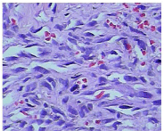
The patient is a 27 years old male with progressive wasting, fatigue and fever for 3 months. A tumor in the right leg was found 1 month ago. On admission, the number of his CD4+ T cells was 13 cells/μl, CD8+ T cells 230 cells/μl, and the CD4+ T cells/CD8+ T cells was 0.01. His WBC was 4.26 × 10⁹/L, Hb was 94 g /L, and platelets were 317 × 109/L. His HIV viral load was 150000 copies/ml. Other biochemical values are: ALT 23 U/L, AST 56 U/L, ALP 274 U/L, γ-GT 234 U/L, total bilirubin 8.7 umol/L, and serum albumin 35.1 g/L. Upon confirmation of Kaposi’s sarcoma pathologically on biopsy sample [1-4], the tumor mass on the leg was surgically removed at Shanghai Public Health Clinical Center (Figure 1).

    Figure 1: Kaposi sarcoma of lower limb in HIV patients, HE x 400 Spindle
cells with dysplasia of allogeneic fibroblasts are cordlike, interlacing, with
unclear cell boundaries, round or spindle nuclei, extending irregularly in all
directions and deeply stained.
The antiretroviral therapy continued and chemotherapy with liposome adriamycin 60 mg was administered after surgical tumor removal for 14 days. The number of CD4+ T cells 7days after the first chemotherapy with liposome adriamycin was 5 cells/μl. The infection around the anus causes ulcers. Defecate more than 10 times a day, it is light yellow and watery. Peripheral anal ulcer gradually increases after local drug exchange treatment (Figure 2A). Bone marrow infusion devices were used for sigmoid colon fistula and intravenous catheterization of sigmoid colon. The perianal biopsy was chronic inflammation with fungal infection. Before the second time chemotherapy again acquisition of autologous bone marrow 50 ml placed bone marrow to save liquid bag, -80°C refrigerator. After 5 days of the second time chemotherapy, autologous bone marrow was transferred back to the implanted bone marrow infusion device. Five courses of chemotherapy were completed in the same way. Primary healing of perianal ulcer after another 5 courses of chemotherapy (Figure 2B). Weight gain is about 6Kg. The number of CD4+ T cells increased to 219 cells/μl. The general condition improved markedly. The second operation closed the sigmoid colon fistula, remanent sigmoid colon back to the abdominal cavity, normal fecal discharge from the anus. Complete healing of ulcer around anus (Figure 2C).

    Figure 1: (A). Kaposi sarcoma patients with anal inflammatory ulcers and fungal infections.
(B). Sigmoid colon fistula, autologous bone marrow preservation before and after chemotherapy, and after six courses of chemotherapy and comprehensive
treatment, the surrounding anal ulcer basically healed.
(C). Closure of sigmoid colostomy and complete healing of ulcer around anus.
Discussion
The pathogenesis and characteristics of Kaposi sarcoma
A rare tumor, first reported by Hungarian dermatologist Kaposi in 1872, is common in Africa and can form endemic in people over the age of 60. Since the 1980s, in Europe and the United States male homosexuals Kaposi’s sarcoma fulminant epidemic, confirmed Kaposi sarcoma has a certain relationship with AIDS, is a kind of common tumor of HIV/AIDS. According to statistics, Kaposi sarcoma is the primary manifestation of HIV infection in about 30% of white gay people. Although Kaposi sarcoma is an aggressive tumor, most patients die from opportunistic infection. Principle of Kaposi sarcoma is still not very clear, may be associated with infection factors, genetic and environmental factors, reports that may be caused by 8 herpes virus, cytomegalovirus infection after the trigger factors. Kaposi sarcoma lesions originated in the middle of the dermis and spread to the epidermis. Histopathology revealed different degrees of fusion between spindle cells and vascular structures. Special dyeing with VIII factor, its cells originated from endothelial cells. Tumor cells are similar to smooth muscle cells, fibroblasts and myoblasts. In the elderly without AIDS, Kaposi sarcoma usually occurs in the toes and legs, presenting as purple or dark brown patches or nodules, mycotic growth or infiltration of soft tissue and invasion of bone tissue. AIDS associated with Kaposi sarcoma, less skin damage or damage is widespread in the skin, mucosa, lymph nodes and internal organs. Generally, the first place found is in the skin of the lower extremity, which is hard knot and slightly uplifted, and gradually expands to the surrounding area, presenting a purple color. The most common site is around the mucosal lymph nodes of the skin and upper digestive tract, which gradually invade the liver, spleen, intestine, brain, lungs, pancreas and testicles. However, the metastatic pathway of sarcoma in the human body has not been clarified, and some experts believe that Kaposi sarcoma is non-metastatic. Once kaposi’s sarcoma occurs, it usually takes 1 year to 1.5 years to die. Concurrent opportunistic infections can only hasten death. In this case, the CD4+T cell number cell 13/μl at the time of kabosi sarcoma was found to be associated with fungal infection and non-tuberculous mycobacterium infection. Chronic inflammatory ulcers around the anus, more than 10 times a day watery yellowish stool, fever and progressive wasting. The number of CD4+ T cells was 5 cells/μl. Continuing this type of chemotherapy could be life-threatening.
Therapeutic ideas and innovations
Chemotherapy may kill tumor cells of Kaposi sarcoma, and combined chemotherapy (such as vinblastine, bleomycin, adriamycin) is effective for Kaposi sarcoma. Liposome adriamycin has a better effect. Liposome is similar to the biological membrane structure of the double molecular utricle, is a single or multiple double lecithin membrane vesicles, its main ingredients are phospholipids, a part of the phospholipid molecules containing phosphate groups with strong polarity (hydrophilic), hydrocarbon chain with nonpolar (hydrophobic). It has the following advantages:
(1) In vivo can be biodegradable, less immunogenicity. (2) Potassium hydrolysate and fat-soluble drugs can be packed and transported, and the drugs are released slowly, and their efficacy lasts a long time. (3) As a result, liposomes can deliver drugs directly to cells, avoiding the use of high concentrations of free drugs and thus reducing adverse reactions. (4) normal tissue capillary wall is complete, most of the liposome can’t penetrate, and capillary permeability increase tumor growth part, increase the amount of liposomes doxorubicin gathered, and due to the slow release of adriamycin, applied directly to the tumor site, increase the therapeutic effect.
But chemotherapy may exacerbate immune damage, aggravate opportunistic infections and endanger lives. In this case, the patient already had chronic inflammatory ulcers around the anus, which were aggravated after a chemotherapy. We use sigmoid colostomy to make feces not pass through the rectum and reduce the stimulation of anal contamination. During the operation of sigmoid colon fistula, the infusion port was buried under the skin of the left lower abdomen through the sigmoid colon vein intubation. It can be placed before chemotherapy acquisition of autologous bone marrow -80°C refrigerator. Three days after the end of chemotherapy, chemotherapy drugs have been basically eliminated from the body. At this time, we were buried in the left lower abdomen hypodermic infusion port after thawing the preserved bone marrow into the portal vein.
We have performed splenectomy plus transomentum right venous catheterization in patients with liver cirrhosis and hypersplenism during AIDS with decompensated stage, and buried the infusion port under the upper abdomen. After surgery, the puncture was embedded in the upper abdomen infusion port, and the autologous bone marrow was injected into the liver through the right omentum vein and portal vein. The results showed that the liver function improved significantly and the original severe cirrhosis was reversed to mild cirrhosis. In addition, during the follow-up, the patient’s CD4+T lymphocytes were found to be significantly increased, which promoted the immune reconstruction. In HIV/AIDS patients with cirrhosis after splenectomy, neutrophils, because of the rapid increase in red blood cell and platelet function after splenectomy, places to reduce damage to these cells, and normal bone marrow hematopoietic function, make the blood cells increased rapidly. The hematopoietic stem cells in the bone marrow differentiate into lymphoid stem cells and then into precursor T cells. T cells need to enter the thymus to mature into T lymphocytes. Since the adult thymus has shrunk, T lymphocytes should not generally increase rapidly. Given that the liver has hematopoietic function in the embryonic stage, we hypothesized that the autologous bone marrow hematopoietic stem cells were injected into the liver, which, under the action of certain cytokines, promoted the development of T cells in the anterior thymus. So if this hypothesis is true, autogenous bone marrow transfusion via portal vein should also promote immune reconstruction in AIDS patients without cirrhosis [5-10].
We removed Kaposi’s sarcoma in this patient, continued antiretroviral therapy, nutritional support therapy and chemotherapy. The patient’s CD4+ T lymphocytes increased from 5 cell/μl before the second chemotherapy, after which the autologous bone marrow was injected through the portal vein, and after another 5 chemotherapy, CD4T lymphocytes increased to 219 cells /μl, significantly promoting immune reconstruction. HIV was not detected in the blood after treatment with antiretroviral therapy. Once again, we closed the sigmoid colostomy and completely healed the ulcer around the anus.
References
- Mani D, Neil N, Israel R, Aboulafia Dm. A retrospective analysis of AIDSassociated Kaposi’s sarcoma in patients with undetectable HIV viral loads and CD4 counts greater than 300 cells/mm3. J Int Assoc Physicians AIDS Care (Chic). 2010; 9: 73.
- Pyakurel P, Pak F, Mwakigonja AR, kaaya E, Biberfeld P. KSHV/HHV-8 and HIV infection in Kaposi’s sarcoma development. Infect Agent Cancer. 2007; 2: 4.
- Azzi S, Smith SS, Dwyer J. YGLF motif in the Kaposi sarcoma herpes virus G-protein-coupled receptor adjusts NF-κB activation and paracrine actions. Oncogene. 2014; 33: 5609-5618.
- Chu K, Misinde D, Massaquoi M, Pasulani O, Mwagomba B, Ford N, et al. Risk factors for mortality in AIDS-associated Kaposi sarcoma in a primary care antiretroviral treatment program in Malawi. Int Health. 2010; 2: 99-102.
- Mark H, Stephen S, Alex T, Giguere P, Barrett L, Haider S, et al. CIHR Canadian HIV Trials Network Coinfection and Concurrent Diseases Core Research Group: 2016 Updated Canadian HIV/Hepatitis C Adult Guidelines for Management and Treatment. Can J Infect Dis Med Microbiol. 2016; 2016: 4385643.
- Deneve JL, Shantha JG, Page AJ, Wyrzykowski AD, Rozycki GS, Feliciano DV. CD4 count is predictive of outcome in HIV-positive patients undergoing abdominal operations. The American Journal of Surgery. 2010; 200: 694-700.
- Feng T, Feng X, Jiang C, Huang C, Liu B. Sepsis risk factors associated with HIV-1 patients undergoing surgery. Emerging Microbes and Infections. 2015; 4: e59.
- Liu B, Chen X, Shi Y. Curative Effect of Hepatic Portal Venous Administration of Autologous Bone Marrow in AIDS Patients with Decompensated Liver Cirrhosis. Cell Death & Disease. 2013; 4: e739.
- Moskowitz AJ, Yahalom J, Kewalramani T, Maragulia JC, Vanak JM, Zelentz AD, et al. Pretransplantation functional imaging predicts outcome following autologous stem cell transplantation for relapsed and refractory Hodgkin lymphoma. Blood. 2010; 116: 4934-4937.
- Feller L, Khammissa RA, Gugushe TS, Chikte UM, Wood NH, Meyerov R, et al. HIV-associated Kaposi sarcoma in African children. J SADJ. 2010; 65: 20-22.
