
Abstract
Myelodysplastic Syndromes (MDS) is a heterogeneous myeloid clonal disease that originates from hematopoietic stem cells, characterized by abnormal differentiation and development of myeloid cells, and manifested as ineffective hematopoiesis, refractory cytopenia, and hematopoiesis with functional failure, there is a high mortality rate. MDS combined with decompensated cirrhosis is more ineffective with conservative treatment. One case of MDS combined with decompensated cirrhosis was observed in our hospital, the platelet counts were 2.5-3×10⁹/L before surgery. The splenectomy combined with autologous bone marrow transhepatic intrahepatic infusion was performed. It was found that after a brief rebound of platelets day 1 after surgery, the platelets decreased to 5×10⁹/L day 10 after surgery, and the autologous bone marrow was infused through the right vein of the omentum by day10, day 30, day 60, and day 90 after surgery, and the platelets gradually increased by 55×109/L by day 10, and the supplement of platelet therapy was not needed, liver function returned to normal, liver CT volume returned to normal, ascites disappeared. One year follow-up, the platelet count was 50×109/L, bone marrow smear can see platelets and no megakaryocytes. After 3-year follow-up, the platelets count was 38×109/L, and liver function was still normal and the general condition of this patient was good. Autologous bone marrow infusion can improve the clinical symptoms of MDS with decompensated cirrhosis, especially the recovery of platelets, no megakaryocytes was seen in the bone marrow, and we speculate that the possibility of platelet production in the liver needs further confirmation by experiments.
Keywords: Myelodysplastic syndrome; Cirrhosis; Hemorrhage; Autologous Bone Marrow; Splenectomy
Introduction
Myelodysplastic Syndromes (MDS) are a heterogeneous myeloid clonal disease that originates from hematopoietic stem cells and is characterized by abnormal differentiation and development of myeloid cells. Often, it is characterized by ineffective hematopoiesis, refractory cytopenia, hematopoietic failure, and a high mortality rate. The treatment of decompensated cirrhosis is currently a difficult problem in medicine. If MDS is combined with decompensated liver cirrhosis, conventional conservative treatment is less effective. We used splenectomy plus autologous bone marrow transhepatic intrahepatic infusion to treat one case of MDS with decompensated cirrhosis, and received good results.
Case Presentation
A 63-year-old man presented to the department with 20-year chronic hepatitis B history. He had abdominal distension, fatigue, gums, repeated subcutaneous hemorrhage, and blood stasis over 12 months. In March 2014, the liver cirrhosis in decompensation stage combined with the MDS in surgical ward. The patient was infected with hepatitis B for 20 years ago due to abdominal distension and fatigue. He was treated with general liver-protecting drugs. One year ago, there was abdominal distension, fatigue and bleeding gums, lamivudine, liver-protecting drugs, ronallide and flumetimilis was treated. 5 months ago, gums and subcutaneous hemorrhage were obvious, platelets were as low as 2.5~3×10⁹/L, and intermittent platelet transfusion was used. After one unit of platelet transfusion, each time it could reach 20~25×109/L. After 10 days, the platelet was reduced to 2.5~3×10⁹/L, and the platelet was infused every 7~10 days.
Examination after Admission
The whole body skin and sclera had a slight yellow stain, the gums had bleeding, and there were scattered blood spots under the skin with no spiders. There was no abnormality in the chest. The abdomen was flat, the spleen could reach 3cm below the left costal margin, and the abdomen had a mobile dullness. Blood routine examination: White Blood Cell (WBC) was 6.25×10⁹/L, Hemoglobin (Hb) was 91g/L, platelet was 24×109 /L, liver function: ALT 33U/L, AST 56U/L, total bilirubin 63.7umol/L, direct bilirubin 37.2umol/L, albumin 46g/L. Abdominal Computed Tomography (CT) examination showed cirrhosis, small liver volume, ascites around the liver, and enlarged spleen.
Treatment Process
On March 7, 2014, splenectomy and perfusion of the right venous catheter were performed under general anesthesia, and the infusion port was embedded in the upper abdomen to establish a percutaneous puncture channel for infusion of fluid to the portal vein. One unit platelet was infused before surgery. The midline incision of the upper abdomen was performed and pale yellow ascites (about 2200mL) was discharged. The splenic artery was first ligated, then the ligament around the spleen was removed and the spleen was removed. Small pieces of liver tissue were cut for pathological examination. The blood remaining in the spleen and the oozing blood were about 1200mL, and were washed back from the peripheral vein after washing through the blood cell return system. The operation was very successful. Blood routine was reviewed by day 1 after surgery, with WBC 40.69×10⁹/L, Hb 104g/L, platelets 68×109/L. Blood routine was reviewed by day 10 after surgery, with WBC 16.42×10⁹/L, Hb 103g/L, and platelets 5.2×10⁹/L. One unit platelet was infused again, and 20 ml autologous bone marrow was collected and infused through the subcutaneous infusion port. Blood routines were reviewed by day 30 after surgery, with WBC 14.12×10⁹/L, Hb 96g/L, and platelets 8×10⁹/L. Liver function: ALT 28U/L, AST 37U/L, total bilirubin 36.3umol/L, direct bilirubin 12.4umol/L, albumin 38.7g/L. Ascites basically disappeared; the diuretic gradually reduced and then was stopped. 20mL autologous bone marrow was infused through the infused port. Blood routine examination was reviewed by day 60 after surgery, with WBC 12.12×10⁹/L, Hb 98g/L, platelet 14×109/L, liver function: ALT 20U/L, AST 37U/L, total bilirubin 19.3umol/L, direct bilirubin 9.4umol/L, albumin 41.7g/L. 20ml autologous bone marrow was infused from the infusion port under the skin. Blood routine examination was performed by day 90 after surgery, with WBC 14.12×10⁹/L, Hb 124g/L, platelet 28×109/L, liver function: ALT 34U/L, AST 37U/L, total bilirubin 11.8umol/L, direct bilirubin 5.5umol/L, albumin 40.9g/L. For the fourth time, 20ml autologous bone marrow was infused through a subcutaneous infusion port. The abdominal CT examination revealed liver cirrhosis, the liver volume was slightly increased compared with that before surgery. There was no ascites around the liver and the spleen was missing (Figure 1). Platelets reached 55×109/L. The body weight gained 8kg in 6 months after surgery. After the diuretic was stopped, there was still no ascites. There was no spontaneous bleeding after stopping the platelet transfusion. The general condition has improved significantly. One year follow-up after surgery, liver function was normal, and platelets were 50×109/L. Bone marrow smear examination, bone marrow classification was more common in mature cells, mononuclear, abnormal lymphocytes were easy to see, no megakaryocytes were seen in the whole film, and platelets were visible. Three years after the operation, the liver function was normal and the platelets were 38×109/L. The general situation is still good.
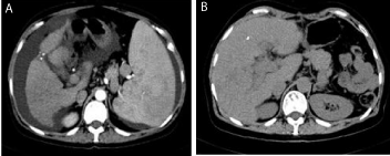
    Figure 1: CT findings of liver before and after treatment.
A: There is ascites around the liver showing the upper kidney superior plane
before surgery, and the liver volume is reduced due to cirrhosis. Spleen
enlargement will push the left kidney down and the left kidney will not be seen
on the CT image.
B: After splenectomy + autologous bone marrow transfusion through the
portal vein after one year, the liver volume increased significantly. Imaging
CT showed that left kidney was seen in the plane of the upper kidney.
Discussion
Transplantation of mesenchymal stem cells for the treatment of post-hepatitis cirrhosis, autoimmune liver disease, alcoholic cirrhosis, schistosomiasis cirrhosis, and so no, has a certain efficacy [1-4]. In this decompensated cirrhosis patient with MDS, we performed splenectomy with autologous bone marrow transfusion via portal vein in the same manner. The basis platelet was 24×109/L, and one unit platelet was infused before surgery. The splenic artery was firstly isolated and ligated, and large amounts of platelets stored in the spleen were introduced into the portal vein through the splenic vein. The huge spleen was taken out during the operation, and the residual blood of the spleen and the oozing blood of the wound were washed by the blood cell returning device and then returned to the surrounding vein. Therefore, the platelets increased significantly on the first day after surgery, and reached 68×109/L. Due to the abnormal clonal hematopoiesis of the megakaryocyte system in the bone marrow, platelets were hardly produced, so the platelets were reduced to 5.2×10⁹/L by day 10 after surgery. One unit platelets was infused again, and the bone marrow was infused via infusion channel, which was implanted in operation. Liver function improved by day 30 after surgery. Ascites basically disappeared. The diuretic was gradually reduced, and was stopped by day 30 after surgery. It indicated that autologous bone marrow intrahepatic infusion significantly promoted liver function reconstruction. The clinical observation is that after spleen resection, the blood vessels in the spleen are selfreturned due to ligation of the splenic artery during surgery. The platelets reach 68×109/L on the first day after surgery, and then the platelets fell rapidly due to platelet dysfunction of bone marrow. After autologous bone marrow transfusion through the portal vein, the liver function gradually returned to normal, and the number of platelets gradually increased. After the second autologous bone marrow portal infusion, platelets were not required to be transfused, and spontaneous bleeding disappeared. The platelet count is continuously increased and reached 40×109/L or more. However, the bone marrow smear examination showed that the whole piece could not find megakaryocytes and the platelets were visible. According to current medical knowledge, platelets are derived from megakaryocytes in the bone marrow. After autologous bone marrow was infused through the portal vein, platelets were found in the surrounding blood and bone marrow, but megakaryocytes were not found in the bone marrow smear. Where did the platelets come from? The bone marrow contains mesenchymal stem cells, hematopoietic stem cells, fat cells and various cytokines. The mesenchymal stem cells promote liver function reconstruction [5,6]. The liver has hematopoietic function at the stage of the embryo, after autologous bone marrow infused into the liver, does the hematopoietic stem cells in the bone marrow restore hematopoietic function in the liver? On the other hand, secrete certain cytokines to promote the megakaryocyte series to restore platelet function?
MDS is divided into primary and secondary, the cause is unknown in primary MDS. The secondary MDS is related to longterm chemotherapy, radiotherapy, secondary to tumors, autoimmune diseases, and so no [7]. So MDS had diversified clinical manifestations and lack the special performance, such as anemia, bleeding and infection. Mainly manifested as a decrease in whole blood cells or a significant decrease in a certain lineage of cells, but the proliferation of myeloid cells is active, and mature and naive cells have morphological abnormalities [8], it is pathological hematopoiesis. It is currently believed that this disease occurs as a result of clonal variation in early hematopoietic stem cells and damage. The studies of chromosome banding analysis in bone marrow cells and glucose-6-phosphate dehydrogenase isozyme suggest that MDS is derived from stem cells and a clonal disease [9]. The onset of this disease is more insidious, it is more common in male middle-aged and older people, especially over 50 year old about 70% cases. Whether or not to decrease the function of intracellular repair gene mutation with age may also be one of the pathogenic factors. For unexplained refractory anemia, MDS should be considered in the exclusion of other hematopoietic and non-hematopoietic disorders that can cause cytopenia and pathological hematopoiesis.
General supportive care for MDS includes blood transfusion, erythropoietin, granulocyte colony stimulating factor or granulocytemacrophage colony stimulating factor and they were used for older MDS or low-risk MDS. The main purpose of supporting treatment is to improve the symptoms of MDS, prevent bleeding and infection and improve the quality of life. We injected autologous bone marrow into the decompensated cirrhosis patient with MDS via the portal vein, which not only restored liver function, but also caused the abnormal MDS clinical bleeding symptoms to disappear. Whether the patient’s ancestral hematopoietic stem cells are normal and the differentiation into the megakaryocyte cell line is abnormal, so the bone marrow rarely produces platelets. But the bone marrow is infused into the liver via the portal vein, and the ancestral hematopoietic stem cells promote platelet production in the liver? This provides a meaningful reference for the treatment of other MDS.
Acknowledgement
This work was supported by the special pilot project of the Chinese Academy of Sciences, “Integrated Research on Stem Cell Application Strategy”.
References
- Liu B, Chen X, Wang Y, Shi Y. Curative effect of hepatic portal venous administration of autologous bone marrow in AIDS patients with decompensated liver cirrhosis. Cell Death Dis. 2013; 4: e739.
- Xue R, Meng Q, Dong J, Li J, Yao Q, Zhu Y, et al. Clinical performance of stem cell therapy in patients with acute-on-chronic liver failure: A systematic review and meta-analysis. J Transl Med. 2018; 16: 126.
- Rajaram R, Subramani B, Abdullah B, Mahadeva S: Mesenchymal stem cell therapy for advanced liver cirrhosis: A case report. JGH Open. 2017; 1: 153- 155.
- Faidah M, Noorwali A, Atta H, Ahmed N, Habib H, Damiati L, et al. Mesenchymal stem cell therapy of hepatocellular carcinoma in rats: Detection of cell homing and tumor mass by magnetic resonance imaging using iron oxide nanoparticles. Adv Clin Exp Med. 2017; 26: 1171-1178.
- Jin J, Hu C, Yu M, Chen F, Ye L, Yin X, et al. Prognostic value of isocitrate dehydrogenase mutations in myelodysplastic syndromes: A retrospective cohort study and meta-analysis. Plos One. 2014; 9: e100206.
- Hull M, Shafran S, Wong A, Tseng A, Giguere P, Barrett L, et al. CIHR canadian HIV trials network coinfection and concurrent diseases core research group: 2016 updated canadian HIV/Hepatitis c adult guidelines for management and treatment. Can J Infect Dis Med Microbiol. 2016; 2016: 4385643.
- Lyle L, Hirose A. Iron overload in myelodysplastic syndromes: Pathophysiology, consequences, diagnosis, and treatment. J Adv Pract Oncol. 2018; 9: 392-405.
- Lowenberg B. Introduction to a series of reviews on myelodysplastic syndromes. Blood. 2019.
- Bell JA, Galaznik A, Huelin R, Stokes M, Guo Y, Fram RJ, et al. Systematic literature review of treatment options and clinical outcomes for patients with Higher-Risk myelodysplastic syndromes and chronic myelomonocytic leukemia. Clin Lymphoma Myeloma Leuk. 2018; 18: 157-166.