Abstract
Background: Breast cancer is the most common malignancy diagnosed in female, it was estimated that new cancer cases and cancer deaths were 1.3 million and 327,000 every year. Breast cancer is the most malignant type in females affecting 1 in every 8 female, it affect old age starting from above 50 years old but may affect also young age. Women undergone surgery or neoadjuvant treatment followed by surgery must be under follow up for a long time to detect any recurrence or metastases or even the development of 2nd primary.
Materials and Methods: Retrospective study done at Tanta cancer center and general surgery department, faculty of medicine. Fayoum University between start of 2004-2009.
Results: 170 patients collected between 2004-2009 with the age at diagnosis was 27-71 years and median age is 49 years old of patients, 90 was premenopausal while 80 was postmenopausal patients.
All patients undergone follow up regularly according to schedule by routine visits. Local recurrence detected in 10 cases with liver metastases in 10 cases and pulmonary metastases in 10 cases with 41 patients died by end of 15 years mainly postmenopausal.
Conclusion: The use of regular and closed follow up with definite schedule is of great value for detecting any progress development of local recurrence, distant recurrence or even the development of another primary.
Keywords: Breast Cancer; Follow Up; Outcome; Recurrence
Abbreviations
IDC: Infiltrating Duct Carcinoma; ILC: Infiltrating Lobular Carcinoma; MRM: Modified Radical Mastectomy; CBS: Conservative Breast Surgery; LVI: Lymph Vascular Invasion; PNI: Peri Neural Invasion; LN: Lymph Node; UOQ: Upper Outer Quadrant; LOQ: Lower Outer Quadrant; ER: Estrogen Receptors; PR: Progesterone Receptors; BC: Breast Cancer; DFS: Disease Free Survival; OFS: Overall Free Survival.
Introduction
Breast cancer is the most common malignancy diagnosed in female [1]. Worldwide, it was estimated that new cancer cases and cancer deaths were 1.3 million and 327,000 every year [2].
Breast cancer is a type of cancer that starts in the breast. Cancer starts when cells begin to grow out of control. Breast cancer is the most malignant type in females affecting 1 in every 8 females worldwide, it can affect any age with most of types on old age starting from above 50 years old. Many women are wishing and excited to be finished with breast cancer treatment. But it can also be a time of worry, being afraid of recurrence of cancer again [3].
Women undergone surgery or neoadjuvant treatment followed by surgery must be under follow up for a long time to detect any recurrence or complications or even the development of 2nd primary.
All patients must have scheduled for investigations either in clinical examination or radiological and laboratory investigations [4].
Material and Methods
170 patients that undergone examination and diagnosis at surgical department, Tanta cancer center and General surgery department, Faculty of medicine, Fayoum University, Egypt, in the period of 2004-2009 was retrospectively collected with their full data as regard the age, pre and postmenopausal status, method of diagnosis, type of surgery done, neoadjuvat treatment and adjuvant treatment, hormonal therapy taken post-operative and development of recurrence either loco regional or distant.
All patients collected during this period treated with surgery either by Conservative Breast Surgery (CBS) or Modified Radical Mastectomy (MRM), those undergone neoadjuvant treatment were included in this study with all patients collected followed up for 10-15years with regular follow up by regular visits and whom missed were phoned to evaluate their status, the follow up was by clinical examination by oncology physician with radiological examination and laboratory needed, all patients under gone ultrasound of abdomen and pelvis for all patients with chest X-ray as regular and CT chest when suspected metastasis in X-ray and bone scan only done during follow up for patients with bony pain or complain and mammographic examination done for all patients regularly every 6months during the 1st two years after surgery and adjuvant then annually after that in both patients undergone MRM or CBS together with chest X-ray and Ultrasound abdomen and pelvis .
Results
170 patients was collected on study done at Tanta cancer center and faculty of medicine, Fayoum university between 2004-2009 with the age at diagnosis was 27-71 years and median age is 49 years old of patients, 90/170 ( 53%) was premenopausal while 80/170 (47%) was postmenopausal patients.
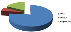
Of those patients all of them undergone mammographic examination with the size of tumor ranged between 2*1cm-5*4cm (all patients was T1&T2), with pathological diagnosis was by FNAC (fine needle aspiration cytology) 130/170 patients (76.48%), true cut biopsy in diagnosis was in 10/170 patients (5.88%) and by lumpectomy 30/170 patients (17.64%) (Figure 1).

Figure 1: Diagnosis of cases.
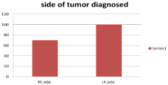
They were diagnosed on Right side breast cancer in 70/170 patients (41.18%) and the rest of patients 100/170 (59.82%) patients diagnosed at Left side (Figure 2).

Figure 2: Sides of tumor diagnosed.
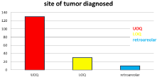
Of them 130/170 (76.48%) tumors located at Upper Outer Quadrant (UOQ) of breast while 10/170 (5.88%) tumors located retro areolar and 30/170 (17.64%) tumors located at Lower Outer Quadrant (LOQ) (Figure 3).

Figure 3: Site of tumor diagnosed.
The least surgical margin was 0.2cm with the largest margin was 3cm and no margin infiltration at all cases but skin fumigation and infiltration was diagnosed in four cases.
During collection of data 10/170 (5.88&) patients was found to have neoadjuvant chemotherapy for down staging before surgery then undergone surgery while 160/170 (94.12%) had surgery from the start.
Most of patients in this study diagnosed as Infiltrating Duct Carcinoma (IDC) 140/170 (82.35%), while 10/170 (5.88%) diagnosed as infilterating lobular carcinoma, 10/170 (5.88%) diagnosed as mixed duct and lobular carcinoma and 10/170 (5.88%) diagnosed as medullary carcinoma of breast cancer type. Carcinoma in Situ (CIS) was found in 20/170 (11.76%) of cases diagnosed in this study (Table 1).
    Type of pathology
    Number of patients
    Percentage %


    IDC
    140
    82.35%


    ILC
    10
    5.88%


    Mixed
    10
    5.88%


    Medullary carcinoma
    10
    5.88%


    CIS
    20
    11.76%


    Stages


    Stage II
    160
    94.12%


    Satge III
    10
    5.88%
Table 1: Types of pathology at diagnosis.
During pathological examination found that most of cases was stage II breast cancer 160/170 (94.11%) while 10/170 diagnosed as stage III breast cancer, with lymph vascular (LVI) and perineural (PNI) invasion detected in only 10/170 cases diagnosed.
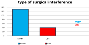
The surgical procedure done as modified radical mastectomy in 130/170 patients (76.48%) and conservative breast surgery with axillary dissection was done in 40/170 patients (23.52%), (Figure 4).

Figure 4: Types of surgical interference.
The largest number of Lymph nodes (LN) harvested during axillary dissection was 11-28 in number while the number of infiltrated nodes was 1-8 of them when 40/170 cases showed capsular invasion of the examined infiltrated Lymph node.
On biological examinations, ER (Estrogen Receptors) was +ve in 130/170 (%) and –ve in 40/170 (%), while PR (Progesterone Receptors) found as +ve in 130/170 cases (%) and –ve in 40/170 cases also (%), HER/2NEU (Herceptin) was found as +ve in 20/170 cases (%) and –ve in 150/170 cases (%) (Table 2).
    Biological marker
    Number of patients


    ER


    +ve
    130


    -ve
    40


    PR


    +ve
    130


    -ve
    40


    Her/2neu


    +ve
    20


    -ve
    150


    Tripple –ve
    40
Table 2: Biological results of tumors.
Multicentericity was present in 10/170 patients while multifocality found in 30/170 patients (%) (Table 3).
    Type of tumor detected
    Number of patients
    Percentage


    Unifocal
    140/170
    82.35%


    Multifocal
    10/170
    5.88%


    Multicenteric
    20/170
    11.77%
Table 3: Multifocality and Multicentericity.
All patients was subjected for adjuvant chemotherapy and radiotherapy while only 140/170 patients had hormonal therapy for 5 years after finishing their chemo radiation therapy.
Local recurrence diagnosed in 10/170 patients of them with 2 cases developed post radiotherapy lymphangiosarcoma treated by simple mastectomy after CBS while the remaining 8/10 patients developed local recurrence treated with simple mastectomy while metastasis detected in 10/170 in liver and 10/170 in lung, 4 cases developed cancer ovary after 5-10ys of 1st primary and one case developed synchronous endometrial and ovarian carcinoma 14 years later treated by surgery of pan hysterectomy and chemotherapy and died 15 years from the diagnosis of the breast cancer, and one patient developed pleomorphic sarcoma of the Rt.psoas major muscle with excision and follow up.
DM in 80/170 patients and hypertension in 70/170 while 30/170 had cardiac problem during period of treatment.
Discussion
Breast cancer is the most common type of malignancy in women diagnosed in one every eight (8) women of population. Breast Cancer (BC) has defined as one of the main causes of morbidity and mortality for women. Each year, at least a million women are diagnosed with this disease in the world and the number of deaths is estimated in 300,000 [5]. Annually, 180,000 new cases and 30,000 deaths are reported in the United States [6].
After potentially curative treatment for breast cancer, it is common clinical practice for patients to be followed up for many years. However, controversies surround follow up, and its value is uncertain [7]. International figures suggest that about 30% of women will develop recurrence after treatment for primary breast cancer, figures for early stage disease being lower [8,9]. Today, the combined effects of screening, early detection, and advances in treatment have resulted in increasing numbers of patients diagnosed with breast cancer in follow up clinics.
Whether early detection of loco regional recurrence leads to a survival benefit, however, is still controversial, with the current evidence suggesting that early detection and treatment of asymptomatic loco regional recurrence has no benefit to overall survival compared with treatment of symptomatic recurrence [10- 12].
The aim of follow up is to detect any local recurrence, regional recurrence or distant metastases or the development of 2nd primary either in breast or elsewhere in the body.
This study is retrospective study collected in Tanta Cancer Center and General surgery department, faculty of medicine, Fayom University, EGYPT, at the period between start of 2004 and start of 2009 for females diagnosed with breast cancer.
With current treatment protocols the local recurrence rate is 1%- 2% per year after breast conserving treatment (and radiotherapy) [7], and 1% after mastectomy. The usual treatments for local recurrence surgery and radiotherapy are more effective if used in the earliest phases. Treatment is aimed at maximizing the chance of long-term local control, as the effect of uncontrolled local recurrence on the woman’s quality of life can be substantial [13]. Local recurrences are more commonly diagnosed during routine follow-up at a time when the patient is asymptomatic. The percentage of patients with a recurrence being asymptomatic at time of detection varied between 9% [8] and 52% [15].
Distant recurrence Common presentations in the symptomatic groups include bone pain, shortness of breath, palpable lesions or enlarged lymph nodes [16]. Most patients who are symptomatic present at interval visits [15,17,18] however, some patients wait to attend routine follow-up visits to discuss symptoms [18].
Contralateral breast cancer, new contralateral primaries are usually diagnosed as part of the regular mammographic screening while patients are asymptomatic [19]. The outcomes are based on tumor characteristics and are independent of the original cancer [19- 21].
The specific methods of detection studied include self-detection, clinical examination, mammography and ultrasound. The reported rates that each of these methods contribute to detection varies. Many studies report that for symptomatic recurrences or new cancers, over 50% are detected by the patient [16]. Over half of asymptomatic recurrences were detected by clinical examination [14-16].
Studies found 12% of asymptomatic recurrences were detected by mammography [16,17]. Eighty-three percent of contralateral breast cancers detected by mammography alone had good or excellent prognostic characteristics [22,23]. Studies indicated that patients with contralateral breast cancer detected by mammography had increased overall survival compared to self-detection [24,25]. Studies found patients with local recurrence detected by mammography showed an increased overall survival compared to physical examination [19,26].
No primary studies were identified which addressed the use of PET or MRI in routine follow-up care.
In this study all patients were under regular follow up with regular visits or by phone with the 1st year was one visit every 6 months then annually, with all patients had routine metastatic work up and radiological diagnosis in the form of Ultrasound of abdomen and pelvis and Triphasic CT only reserved for patients had liver metastases, chest X-ray and CT chest in cases suspected to have pulmonary metastasis together with bilateral mammography for patients undergone CBS and unilateral for those had MRM with bone scan only done for patients had bony pain or complain. During treatment of patients, 130/170 (76.47%) treated by Modified Radical Mastectomy (MRM) and 40/170 (23.53%) treated by Conservative Breast Surgery (CBS), of them 10 patients out of 170 (5.88%) had local recurrence 8 patients of them was symptomatic and 2 patients with asymptomatic, and all of them was treated by CBD with no local recurrence after surgical interference by MRM and also all the recurrence in 10 cases detected in premenopausal patient when diagnosed as recurrence and all of them was treated by simple mastectomy, two of them has post irradiation lymphangiosarcoma and still under follow up [27,28].
Some of literatures indicated the association of grade of primary tumor and development of secondary tumor [27].
During follow up, 10 patients out of 170 developed (5.88%) liver metastasis that confirmed by Triphasic CT scan 8 of them was symptomatic and 2 patients was asymptomatic detected during routine follow up and imaging and 10/170 (5.88%) developed pulmonary metastasis, 6 patients was symptomatic of cough and chest pain while 4 patients asymptomatic and detected by radiological imaging that suspected on chest X-ray and then confirmed by CT chest for diagnosis and both of them undergone chemotherapy (Table 4).
    Type of recurrence
    Symptomatic
    Asymptomatic
    Number of cases


    Local    recurrence
    8
    2
    10


    Pulmonary    metastases
    6
    4
    10


    Liver    metastases
    8
    2
    10
Table 4: Recurrence during follow up.
4 patients out of 170 (2.35%) of the study developed cancer ovary in a period of 10-14years of the diagnosis of breast cancer and one patient developed synchronous cancer ovary and endometrial carcinoma after 14 years of breast cancer then had chemotherapy with development of pelvic recurrence in the 1st year after surgery and died, all of them was post-menopausal at the time of diagnosis of the 2nd primary and only one patient developed retroperitoneal pleomorphic sarcoma 10 years after diagnosis of breast cancer and had undergone excision and still under follow up and 2 patients developed contralateral breast cancer (Table 5).
    Types of 2nd tumor
    Number of patients
    Percentage


    Cancer    ovary
    4/170
    2.35%


    Cancer    ovary and endometrial carcinoma
    1/170
    0.59%


    Retroperitoneal    liposarcoma
    1/170
    0.59%


    Post    irradiation lymphangiosarcoma
    2/170
    1.18%


    Contralateral    breast
    2/170
    1.18%
Table 5: Second primary detected in follow up.
During analysis of data of this study, all patients found to have 0-1 node infiltrated at the time of axillary dissection with surgical treatment with no recurrence detected on patients treated by MRM with all cases detected in patients diagnosed at premenopausal age while 2nd primary detected only after MRM patients and in postmenopausal. All the cases had local recurrence and had 2nd primary all had ER and PR ranged from moderate to markedly positive and received hormonal treatment according to protocol of age for 5 years.
At the end of 5 years after surgery and treatment 2/170 died with overall free survival OFS was 98.8% and disease free survival DFS was 92.94% and after 15 years of follow up 41/170 died by the end of 15 years follow up with 75.9% over all free survival OFS and disease free survival DFS was 69.41%.
Conclusion
As treatment for cancer improves, women are living longer and accordingly efforts to improve survivors, quality of life, without negative impact on their time to recurrence or survival. The use of early detection due to regular and closed follow up with definite schedule is of great value for detecting any progress in follow up as the development of local recurrence, distant recurrence or even the development of another primary and the detection of contralateral breast cancer. The recurrence is mostly noted after treatment by CBS than treatment by MRM and in premenopausal then postmenopausal at time of diagnosis of recurrence while the development of 2nd primary is more on post-menopausal patients.
References
- Nelson HD, Zakher B, Cantor A, Griffin J, O’Meara ES, Buist DS, et al. Risk factors for breast cancer for women aged 40 to 49 years: a systematic review and meta-analysis. Ann Intern Med. 2012; 156: 635-648.
- Confortini CC, Krong B. Breast cancer in the global south and the limitations of a biomedical framing: a critical review of the literature. Health Policy Plan. 2015; 30: 1350-1361.
- Jagsi R, King TA, Lehman C, et al. Chapter 79: Malignant tumors of the breast. In: De Vita VT, Lawrence TS, Rosenberg SA, eds. DeVita, Hellman, and Rosenberg’s Cancer: Principles and Practice of Oncology. 11th ed. Philadelphia, Pa: Lippincott Williams & Wilkins. 2019.
- National Comprehensive Cancer Network (NCCN). Practice Guidelines in Oncology: Breast Cancer. Version 2. 2019.
- Pisani P, Parkin DM, Bray F, Ferlay J. Estimates of the worldwide mortality from cancer in 1990. Int J Cancer. 1999; 83: 18-29.
- Holford TR, Roush GC, McKay LA. Trends in female breast cancer in Connecticut and the United States. J Clin Epidemiol. 1991; 44: 29-39.
- National Health and Medical Research Council. Clinical practice guidelines for management of early breast cancer. Canberra: NHMRC. 2001; 97-102.
- Wong WW, Vijayakumar S, Weichselbaum RR. Prognostic indicators in node negative early stage breast cancer. Am J Med. 1992; 92: 539-548.
- Gasparini G, Bevilacqua P, Dal Fior S, Panizzoni GA, Favretto S, Visonà A, et al. Characterization of node negative breast cancer by clinico-pathologic and biologic factors. Anticancer Res. 1990; 10: 205-208.
- te Boekhorst DS, Peer NG, Van der Sluis RF, Wobbes T, Ruers TJ. Periodic follow-up after breast cancer and the effect on survival. Eur J Surg. 2001; 167: 490-496.
- Churn M, Kelly V. Outpatient follow-up after treatment for early breast cancer: updated results after 5 years. Clin Oncol. 2001; 13: 187-194.
- Clemons M, Danson S, Hamilton T, Goss P. Loco regionally recurrent breast cancer: incidence, risk factors and survival. Cancer Treat Rev. 2001; 27: 67- 82.
- National Breast Cancer Centre. Clinical practice guidelines for the management of early breast cancer (2nd Edition). Canberra: Commonwealth of Australia. 2001.
- Perrone MA, Musolino A, Michiara M, Di Blasio B, Bella M, Franciosi V, et al. Early detection of recurrences in the follow-up of primary breast cancer in an asymptomatic or symptomatic phase. Tumori. 2004; 90: 276-279.
- Donnelly J, Mack P, Donaldson LA. Follow-up of breast cancer: time for a new approach? Int J Clin Pract. 2001; 55: 431-433.
- Te Boekhorst DS, Peer NG, Van der Sluis RF, Wobbes T, Ruers TJ. Periodic follow-up after breast cancer and the effect on survival. Eur J Surg. 2001; 167: 490-496.
- Hiramanek N. Breast cancer recurrence: Follow-up after treatment for primary breast cancer. Postgrad Med J. 2004; 80: 172-176.
- Grogan M, Rangan A, Gebski V, Boyages J. The value of follow-up of patients with early breast cancer treated with conservative surgery and radiation therapy. Breast. 2002; 11: 163-169.
- Montgomery DA, Krupa K, Jack WJ, Kerr GR, Kunkler IH, Thomas J, et al. Changing pattern of the detection of loco regional relapse in breast cancer: the Edinburgh experience. Br J Cancer. 2007; 96: 1802-1807.
- Chen C, Orel SG, Hams EE, Hwang WT, Solih LJ. Relation between the method of detection of initial breast carcinoma and the method of detection of subsequent ipsilateral local recurrence and contralateral breast carcinoma. Cancer. 2003; 98: 1596-1602.
- Yau TK, Sze H, Soong IS, Wong W, Chan K, Lau KY, et al. Surveillance mammography after breast conservation therapy in Hong Kong: Effectiveness and feasibility of risk-adapted approach. Breast. 2007.
- National Breast and Ovarian Cancer Centre. Follow-up of patients with early breast cancer: a systematic review. NBOCC, Surry Hills, NSW. 2009.
- Kollias J, Evans AJ, Wilson AR, Ellis IO, Elston CW, Blamey RW. Value of contralateral surveillance mammography for primary breast cancer follow-up. World J Surg. 2000; 24: 983-987; discussion 988-989.
- Robinson A, Speers C, Olivotto I, Chia S. Method of detection of new contralateral primary breast cancer in younger versus older women. Clin Breast Cancer. 2007; 7: 705-709.
- Kaas R, Hart AA, Besnard AP, Peterse JL, Rutgers EJ. Impact of mammographic interval on stage and survival after the diagnosis of contralateral breast cancer. Br J Surg. 2001; 88: 123-127.
- Doyle T, Schultz DJ, Peters C, Harris Z, Solin LJ. Long-term results of local recurrence after breast conservation treatment for invasive breast cancer. Int J Radiat Oncol Biol Phys. 2001; 51: 74-80.
- Storm HH, Jensen OM. Risk of contralateral breast cancer in Denmark 1943- 80. Br J Cancer. 1986; 54: 483-492.
- Ruddy KJ, Partridge AH. Approach to the patient following treatment for breast cancer. Up To Date. 2019.
