Case Report
Austin Therapeutics. 2014;1(1): 3.
Diagnosis of a Unilateral Pulmonary Agenesis in a Term Newborn without Invasive Procedures or Advanced Imaging
Gal Sagie1, Benjamin Z Koplewitz2, Gavri Sagui3, Shlomo Cohen4, Chaim Springer4, Benjamin Bar- Oz5 and Smadar Eventov-Friedman5*
1Department of Pediatrics, Hadassah Ein-Kerem and Hebrew University Medical Center, Israel
2Department of Medical Imaging, Hadassah and Hebrew University Medical Center, Israel
3Department of Pediatric Cardiology, Hadassah and Hebrew University- Medical Center, Israel
4Pediatric Pulmonology Unit, Hadassah and Hebrew University Medical Center, Israel
5Department of Neonatology, Hadassah and Hebrew University -Medical center, Israel
*Corresponding author: Smadar Eventov-Friedman, Department of Neonatology, Hadassah and Hebrew University- Medical Center, Jerusalem, 91120, Israel
Received: August 05, 2014; Accepted: August 12, 2014; Published: August 12, 2014
Abstract
Isolated unilateral pulmonary agenesis is a rare condition. The diagnosis is often missed during infancy and childhood due to non-specific physical findings. We present a term newborn diagnosed with pulmonary artery and ipsilateral lung agenesis using chest radiography and echocardiogram, with no need for invasive procedures or advanced imaging modalities.
Keywords: Pulmonary artery agenesis; Lung hypoplasia; Pulmonary hypertension; Advanced imaging modality
Introduction
Agenesis of a pulmonary artery and hypoplasia/agenesis of the ipsilateral lung in the absence of other congenital cardiac malformations is a rare condition with a prevalence of 1:200,000 in the general population [1]. This congenital defect can be identified incidentally at different ages, including the neonatal period. We report a neonate who had no respiratory symptoms at birth and was diagnosed with agenesis of the left lung and the left pulmonary artery at the age of 5 days. The diagnosis was performed by chest radiograph and was confirmed by detailed color Doppler echocardiography obviating the need for bronchoscopy or CT/MRI studies.
Case Presentation
A-38 week gestation male infant was born vaginally with a birth weight of 3,230 grams. Apgar scores were 9 and 10 at 1 and 5 minutes, respectively. The mother was a 33- year-old white healthy woman. This was her second pregnancy. No medication was taken throughout the pregnancy. Routine ultrasonographic screening examinations uring pregnancy revealed a single umbilical artery and right club foot. The mother chose not to undergo a fetal echocardiogram.
Upon his admission to the nursery, peripheral hematocrit sample was taken due to plethoric skin that showed a borderline high value of 69%. Glucose levels were low (42 mg%) and intravenous glucose infusion was commenced. The physical examination was normal except for right clubfoot. However, there was an impression of decreased sounds of air entry s to the left lung field. No cardiac murmur was heard and peripheral pulses were adequately felt. Club foot was noted at the right side. Blood glucose as well as the hematocrit values normalized shortly after the initiation of the intravenous fluid infusion. A complete blood count showed thrombocytopenia of 78,000. Therefore, a complete sepsis evaluation was preformed and antibiotic treatment was added. After 48 hours mild de-saturation events without tachypnea, dyspnea or grunting were observed and supplemental oxygen (maximal FiO2 -0.3) was occasionally given.
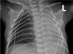
Arterial blood gases were within normal limits. Chest Radiograph (CXR) showed no sign of air or blood vessels in the left chest. A leftward mediastinal shift was seen (Figure 1).

Figure 1: Chest radiograph performed at the age of day 2, mildly rotated to the right side. The left chest is not aerated and the mediastinum is shifted to the left. The left bronchus cannot be identified. Note normal vascularization and aeration of the right lung. Austin Therapeutics 1(1): id1002 (2014) - Page - 02 Smadar Eventov-Friedman Austin Publishing Group Submit your Manuscript | www.austinpublishinggroup.com leftward mediastinal shift was seen (Figure 1).
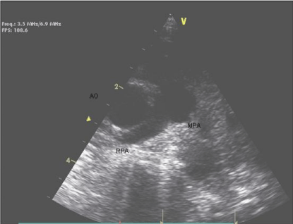
Chest ultrasound showed no sign of air, suggesting no lung tissue in the left thorax. Echocardiogram showed normal cardiac anatomy with agenesis/ hypoplasia of the left pulmonary artery and the left pulmonary veins. Increased flow was noted in the superior vena cava and the right pulmonary vein. No signs of pulmonary hypertension were observed by flow measurement on the tricuspid valve. A small left atrium and a small patent foramen ovale with left to right shunt were seen (Figure 2, clip). Abdominal and brain ultrasound were normal.

Figure 2: Echocardiogram at the age of 2 days. Parasternal short axis showing Aorta (Ao) MPA (Main pulmonary artery) and RPA ( Right Pulmonary Artery ) with no evidence to LPA ( Left Pulmonary Artery).
Within two days the de-saturation events resolved. The repeated blood count was within normal limits. The blood and CSF cultures were negative after 72 hours, at which time the antibiotic treatment was discontinued. The karyotype was normal (46XY). The infant was discharged at the age of 9 days, with his right leg placed in cast for his club foot. Follow-up visits in the pediatric outpatient clinic until the age of one year old revealed normal growth and neurodevelopment. No signs of disease or respiratory distress were found.
Discussion
Developmental anomalies of the lung can be categorized as bronchopulmonary, vascular or combined. Agenesis occurs when a lung lobe, the whole ipsilateral lung, the bronchi and their vessels fail to develop. In aplasia, there is no development of lung or blood vessels, but a rudimentary bronchus can be found. In hypoplasia, a rudimentary bronchus and hypoplastic lung parenchyma can be detected [2-4].
Unilateral pulmonary artery agenesis is a rare condition. It was first described in 1673 [5]. The true incidence of this anomaly is not precisely known. Pulmonary hypoplasia spectrum is reported in 7-26 % of all neonatal autopsies [6]. It occasionally occurs as an isolated lesion but can be associated with a spectrum of skeletal anomalies [7], ipsilateral malformations [8] and congenital cardiac anomalies [9- 11]. The pathogenesis remains unknown, though vascular disruption and teratogenic insults have been considered.
The diagnosis of isolated pulmonary agenesis is often missed during infancy and childhood. Patients may be asymptomatic even throughout their entire lives [1,9,12]. Presenting symptoms might be decreased exercise tolerance and mild dyspnea caused by pulmonary hypertension and congestive heart failure. Recurrent pulmonary infections or hemoptysis and pulmonary edema are less common [9].
Video
Please download Video here
In this reported case, the only presenting sign were mild decreased air entry upon auscultation to the left chest on physical examination and few mild desaturation events in the second day of life. This served as the initial clue to the diagnosis a unilateral pulmonary artery agenesis with ipsilateral lung hypoplasia that was established by chest radiographic and the echocardiographic findings. The uneventful clinical course and the normal development rendered further investigations unnecessary. In our opinion, invasive procedures such as bronchoscopy or cardiac catheterization may be postponed as long as there is no echocardiographic evidence of pulmonary hypertension, and advanced chest imaging such as CT or MRI may not be required at an early stage of life in this condition.
Established facts
Unilateral pulmonary agenesis is a rare condition. The diagnosis usually requires invasive procedures or advanced imaging modality.
Novel insights
The diagnosis of isolated pulmonary lung and artery can be established on chest radiography and echocardiogram.
References
- Bouros D, Pare P, Panagou P, Tsintiris K, Siafakas N. The varied manifestation of pulmonary artery agenesis in adulthood. Chest. 1995; 108: 670-676.
- DeLorimier AA. Congenital malformations and neonatal problems of the respiratory tree. 4th edn. Welach KJ, Randolph JG, Ravitch MM, et al. In: Pediatric surgery. Chicago: year book medical publishers. 1986; 631-44.
- Skandalakis JE, Grey SW, Symbas P. The trachea and lungs. 2nd edn. Skandalakis JE, Grey SW, Symbas P. In: Embryology for surgeons. Baltimore: Williams & Wilkins. 1994; 414-450.
- Nazir Z, Qazi SH, Ahmed N, Atiq M, Billoo AG. Pulmonary agenesis--vascular airway compression and gastroesophageal reflux influence outcome. J Pediatr Surg. 2006; 41: 1165-1169.
- Evrard V, Ceulemans J, Coosemans W, De Baere T, De Leyn P, Deneffe G, et al. Congenital parenchymatous malformations of the lung. World J Surg. 1999; 23: 1123-1132.
- Abrams ME, Ackerman VL, Engle WA. Primary unilateral pulmonary hypoplasia: Neonate through early childhood- case report, radiographic diagnosos and review of the literature. J Perinatol. 2004; 24: 667-670.
- Osborne J, Masel J, McCredie J. A spectrum of skeletal anomalies associated with pulmonary agenesis: possible neural crest injuries. Pediatr Radiol. 1989; 19: 425-432.
- Cunningham ML, Mann N. Pulmonary agenesis: a predictor of ipsilateral malformations. Am J Med Genet. 1997; 70: 391-398.
- Ten Harkel AD, Blom NA, Ottenkamp J. Isolated unilateral absence of a pulmonary artery: a case report and review of the literature. Chest. 2002; 122: 1471-1477.
- Kucera V, Fiser B, Tuma S, Hucin B. Unilateral absence of pulmonary artery: a report on 19 selected clinical cases. Thorac Cardiovasc Surg. 1982; 30: 152-158.
- Vitiello R, Pisanti C, Pisanti A, Silberbach M. Association of pulmonary artery agenesis and hypoplasia of the lung. Pediatr Pulmonol. 2006; 41: 897-899.
- Thomas RJ, Lathif HC, Sen S, Zachariah N, Chacko J. Varied presentations of unilateral lung hypoplasia and agenesis: a report of four cases. Pediatr Surg Int. 1998; 14: 94-95.