Special Article - Red Blood Cells
Thromb Haemost Res. 2019; 3(3): 1030.
Early Candida parapsilosis Sepsis Detection Using Sysmex XN-3000 Hematological Analyzer: Case Report
De Azambuja AP*, Spiri BS, Ferreira D, Trenenpohl J, Bonfim CMS and Comar SR
Universidade Federal do Paraná, Brazil
*Corresponding author: Ana Paula de Azambuja, Universidade Federal do Paraná, Brazil
Received: September 03, 2019; Accepted: October 03, 2019; Published: October 10, 2019
Abstract
Candida parapsilosis sepsis is a rare but fatal disease affecting all age groups, commonly caused by fungal surface contamination of medical devices such as central venous catheters [1]. These infections are relevant and increasing problem in debilitated and immunosuppressed patients, which can evolve from a superficial non-life-threatening disease to a clinical picture of severe sepsis with multiorgan dissemination [2].
An etiological diagnosis of catheter-related candidemia requires the yeasts isolation from both peripheral and catheter blood samples and takes three or more days in obtaining definite culture results, so the average time before an etiological therapy can be administered usually involves waiting more than 98 h, depending on the species of Candida [2].
We describe a young child case where leukogram scatter diagram in XN- 3000 hematology analyzer (Sysmex, Japan) allowed viewing of a distinct blue cloud in erytroblast canal (NRBC), allowing clinicians to start empiric antifungal therapy long before the blood culture results were available.
Case Report
Male, 4 months, diagnosed with Severe Combined Immunodeficiency Syndrome (SCID) due to recurrent infections, perianal dermatitis and disseminated hyperchromic skin lesions, with undetectable TRECs and Krec’s and flow cytometry with T+, Band NK+ lymphocytes, absence of naïve LT (CD45RA) and increased memory LT (CD45RO). The etiological diagnosis of Ommen’s Syndrome was done and the patient received haploidentical Hematopoietic Stem Cell Transplantation (HSCT) with donor father.
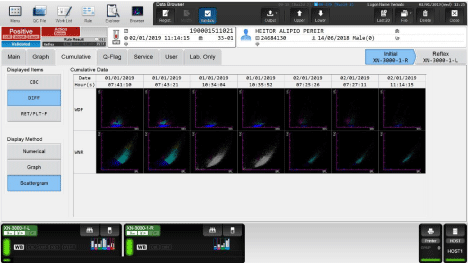
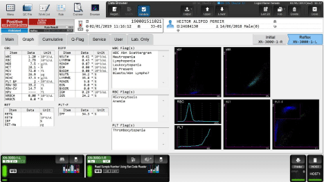
In eleven day after bone marrow transplantation, a routine complete blood count and white blood cell differentials performed on XN-3000 hematology analyzer (Sysmex, Japan) showed a blue cloud in the erythroblast canal (NRBC) suspected of Candida sp. in the dispersion diagram of the leukogram (Figure 1 and 2). Microscopic review of May-Grünwald- Giemsa stained peripheral blood smears performed by experienced hematologists and microbiologist and a positive blood culture done in Bactec FX Top (Becton Dickinson) increased suspicion for candidemia. The agar chocolate culture for identification of the species (VITEK - bioMérieux Brasil), confirmed the presence of C. parapsilosis.

    Figure 1: Dispersion diagram of the leukogram (FS/SS graph) showing a blue cloud in the erythroblast canal (NRBC) suspected of Candida sp. in a routine
complete blood count and white blood cell differentials performed on XN-3000 hematology analyzer.

Figure 2: Cumulative differencial scattergram showing the erythroblast canal (NRBC) suspected of Candida sp.
Thus, although asymptomatic, an early diagnosis of invasive fungal infection was done and started antifungal therapy with Amphotericin B, followed by lock therapy, what was maintained for 14 days after culture negativity. In parallel to use antifungal agent, the Hickmann catheter was changed and three mother donor granulocyte infusions were performed.
Despite to the fact that patient persisted for a long time with febrile neutropenia, no evidence of another infectious disease was found. Patient also presented BCG infection and pulmonary mycobacteriosis, acute grade I Graft versus Host Diesease, which were managed as usual in the HSCT scenario.
Discussion
The detection of Candida species in peripheral blood smears suggests advanced disseminated infections with uncontrollable complications, whether they were detectable or not in clinical examinations, caused by the burden of Candida species in the blood, which is indicative of poor prognosis. In case of candidemia, an etiological treatment must be started only after a positive blood culture. As microbiological testing requires some days to be fully performed and sepsis mortality is increased by any delay in diagnosis and treatment, in some patients the benefit of starting an early empiric antifungal treatment based on the available clinical and laboratory data often outweigh any other risk.
La Gioia et al. described two cases were the presence of distinctive alterations in the 3D-DIFF cytograms could be characteristic of the presence of Candida sp. in the sample. They presented blood cell count performed on BC-6800 hematological analyzer (Mindray, Shenzhen, China). In hematological analyzers, the circulating leukocytes as well as erythroblasts are evaluated by side and forward laser scatter (SS and FS) and by Fluorescence (FL). The resulting Three-Dimensional (3D) scattergram can be rotated to allow a detailed observation of cell population clusters, their complexity, size and nucleic acids cell content [2]. Small size and low internal complexity form a plankshaped cloud, suggestive of yeast infestation, regardless of number. Besides that, neutrophil population may present extension in FC/SS axis, evidencing increase in the size and internal complexity, which occurs when there is yeast phagocytosis by neutrophils. The prompt microscopic review can confirm the presence of circulating fungal pathogen.
In the present case, the viewed of a blue cloud in NRBC channel different from the common pattern found for these cells on the XN-3000 hematology analyzer cytogram allowed us to suspect the presence of Candida sp. in the sample, later confirmed by blood culture. This fact allow early iniciation of antifungal therapy and probably, prevented baby´s death.
Conclusion
Our observations suggest that NRBC cytograms generated by the automatic analyzers may be useful for early diagnosis of Candida sp., enabling to anticipate antifungal therapy, e possible reducing mortality rate of immunosuppressed and HSC transplanted patients.
References
- Devrim I, Yaman Y, Demirag B, Oymak Y, Carti Ö, Özek G, et al. A single center’s experience with Candida parapsilosis related long-term central venous access device infections: the port removal decision and its outcomes. Pediatr Hematol Oncol. 2014; 31: 435–441.
- Hirai Y, Asahata S, Ainoda Y, Fujita T, Miura H, Hizuka N, et al. Candidemia diagnosed from peripheral blood smear: case report and review of literature 1954–2013. Mycopathologia 2015; 180: 111–116.
- La Gioia A, Bombara M, Fiorini F, Dell’Amico M, Devito A, Isola P, et al. Earlier detection of sepsis by Candida parapsilosis using three-dimensional cytographic anomalies on the Mindray BC-6800 hematological analyzer. Clin Chem Lab Med. 2016; 54: e239–e242.