Case Presentation
Annals Thyroid Res. 2019; 5(1): 196-197.
Thyrotoxicosis due to Thyroid Hematoma Precipitated by Anticoagulation Therapy
Asbar H*, Askaoui S, Rafi S, El Mghari G and El Ansari N
1Department of Endocrinology, Cadi Ayyad University, Morocco
*Corresponding author: Asbar H, Department of Endocrinology, Diabetes, Metabolic diseases and Nutrition, Mohammed VI University hospital, Cadi Ayyad University, Marrakesh, Morocco. Portes de Marrakech, Tranche 8 B, villa 37, Marrakech, Morocco
Received: March 14, 2019; Accepted: April 24, 2019; Published: May 01, 2019
Abstract
Intrathyroidal bleeding of non-traumatic origin is of rare occurrence. It can be increased by the use of anticoagulant agents. We report the case of an elderly woman with history of nontoxic multinodular goiter that developed a spontaneous thyroid hemorrhage precipitated by the use of oral anticoagulants (acenocoumarol) for atrial fibrillation. She presented with compressive signs (dyspnea, dysphagia and dysphonia). Cervical computed tomography showed enlarged, heterogeneous thyroid gland, with diffuse infiltration of thyroid lodge with collections measuring for the most voluminous 50x70mm, pushing trachea but that remains permeable. Biological findings revealed increased levels of thyroxine and triiodothyronine with a decreased levels of thyroidstimulating hormone. She was treated with steroids, antibiotics and antithyroid medication in the face of a destructive hyperthyroidism. Evolution was good and a total throidectomy was performed after a month. Only few cases of non traumatic thyroid hemorrhage are described in the literature. It is a potentially life-threatening condition by compression of aerodigestive tract and adjacent vascular axes. Our report is to our knowledge the first case of intrathyroidal bleeding due to acenocoumarol causing transient thyrotoxicosis. It illustrates a rare but potentially lethal complication of anticoagulants especially in elderly population.
Keywords: Thyroid gland; Hemorrhage; Anticoagulation; Thyrotoxicosis
Abbreviations
CT: Cervical Computed Tomography; T4: Thyroxine; T3: Triiodothyronine; TSH: Thyroid-Stimulating Hormone; INR: International Normalized Ratio
Introduction
Spontaneous thyroid hematoma is a rare occurrence that can be favored by anticoagulation therapy. Any neck swelling can be life-threatening by compression of the upper aero digestive tract and adjacent vascular axes. We report the case of a thyroid hematoma revealed by compressive signs in a patient with a history of a nontoxic multinodular goiter treated with vitamin K antagonist (acenocoumarol) for atrial fibrillation. Subsequent course was marked by installation of hyperthyroidism of destructive mechanism following the thyroid hematoma.
Case Presentation
We report the case of an 80-year-old patient with a history of nontoxic multinodular goiter, recently started on vitamin K antagonists (acenocoumarol) for atrial fibrillation, who presented to the emergency department for a progressively increasing neck swelling, causing mild sore throat, breathing difficulty and dysphagia to solids. She also complained of palpitations and a change in her voice.
On examination the patient was asthenic, pale, dyspneic with respiratory rate of 32 cycles/minutes, tachycardia of 120 beats/minutes, blood pressure of 100/60mmHg, and oxygen saturation of 99% under four liters of oxygen. There was a diffuse anterior neck swelling of approximately 08 x 07cm that was very painful on palpation, firm and mobile. The presence of extensive spontaneous bruising in both arms was suggestive of vitamin k antagonist intoxication.
Laboratory findings showed INR of 7 (therapeutic goal for atrial fibrillation 2.0-3.0) with poorly tolerated 6g/dl anemia. TSH level was low at 0.02mIU/L, with an elevated free T4 level at 40pmol/l indicative of hyperthyroidism. Leukocyte count was at 13 400/mm3 predominantly neutrophils with an elevated C-reactive protein at 250mg/L. Blood electrolytes, renal and liver function were normal.
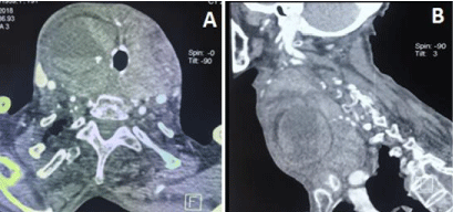
Urgent CT of the neck was done, which showed an enlarged, heterogeneous thyroid gland, with diffuse infiltration of thyroid lodge with collections measuring for the most voluminous 50x70mm (Figure 1). Treatment was based on frozen fresh plasma and red blood cells transfusion with discontinuation of anticoagulation therapy, oxygen therapy, intravenous steroids and antibiotics. Antithyroid medications were initiated for control of increase sympathetic activity before planning surgery. Total thyroidectomy was perfomed after a month with a good evolution.

    Figure 1: (A) Axial section injected CT showing a predominantly liquid
thyroid collection pushing the axis aero-digestive and vasculo-nervous but
that remain permeable. (B) Sagittal section CT showing a large nodular goiter
associated with a large infiltration of the thyroid compartment with poorly
limited collections.
Discussion
Even though thyroid is a highly vascularized gland, thyroid hemorrhage is rare. Recent data from the literature report only few cases of cervical spontaneous hematomas, almost half of which are precipitated by anticoagulation therapy (heparin or vitamin k antagonists for various indications). Other causes include in most cases blunt trauma or fine needle aspiration [1].
Pre-existing thyroid abnormalities, such as the presence of goiter or thyroid nodules, may increase the risk of bleeding. Different mechanisms are proposed such as abnormal thyroid vascular anatomy, and arteriovenous shunting in a cyst or nodule [2,3]. Physical exertion, coughing, straining, or any valsalva maneuver can increase the venous pressure and cause rupture of the vessels resulting in a thyroid hematoma [4].
With the aging of the population, prevalence of goiter is increasing in the elderly population. Multinodular goiter and grave’s disease are the main etiologies of goiter in patients aged 55 years and older [5]. Indications for chronic anticoagulation are also frequent, and can increase risk of bleeding. Different anticoagulation agents can be prescribed (acenocoumarol, warfarin…). Only 3 cases of spontaneous thyroid hemorrhage were reported in patients on anticoagulation with warfarin for atrial fibrillation [1,6,7]. To our knowledge no case of spontaneous thyroid hematoma on acenocoumarol for this indication was reported.
Hyperthyroidism due to follicular tissue destruction leading to release of stored thyroid hormone can be associated with radioiodine therapy, amiodarone-induced thyroiditis as well as lymphocytic thyroiditis, subacute granulomatous thyroiditis, and postpartum thyroiditis [8]. In our case, it was associated to hemorrhage into thyroid nodules which remains rarely reported in the literature [8- 11].
Management of such cases is based primarily on maintaining airway patency. Bleeding within a goiter causing airway compressing requires urgent intubation and surgical evacuation if the patient is instable. In our case, the patient initially refused any surgery and she was stabilized under symptomatic treatment. Conservative treatment is efficient when collections are mild and in an early stage. Acute hyperthyroidism is usually transient and patients may require partial or total thyroidectomy [12].
Conclusion
The use of anticoagulant treatments is becoming more common. Bleeding of thyroid origin is rare but could be favored by the preexisting goiter or nodules. Transient thyrotoxicosis on thyroid hemorrhage is an interesting and rare association. This complication of anticoagulants, especially in geriatric patients, deserves to be known because it can be life-threatening.
References
- Gunasekaran K, Rudd K, Murthi S, Kaatz S, Lone N. Spontaneous thyroid hemorrhage on chronic anticoagulation therapy. Clinics and practice. 2017; 7: 932.
- Vijapurapu R, Kaur K, Crooks NH. A case of airway obstruction secondary to acute haemorrhage into a benign thyroid cyst. Case Rep Crit Care. 2014; 2014: 372369.
- Testini M, Logoluso F, Lissidini G, Gurrado A, Campobasso G, Cortese R, et al. Emergency total thyroidectomy due to non traumatic disease. Experience of a surgical unit and literature review. World J Emerg Surg. 2012; 7: 9.
- Lee JK, Lee DH, Cho SW, Lim SC. Acute airway obstruction by spontaneous haemorrhage into thyroid nodule. Indian J Otolaryngol Head Neck Surg. 2011; 63: 387–389.
- Diez J. Goiter in adult patients aged 55 years and older: etiology and clinical features in 634 patients. J Gerontol A Biol Sci Med Sci. 2005; 60, 7: 920-923.
- Olchovsky D, Pines A, Zwas ST, Itzchak Y, Halkin H. Apathetic thyrotoxicosis due to hemorrhage into a hyperfunctioning thyroid nodule after excessive anticoagulation. South Med J. 1985; 78: 609-611.
- Kokatnur L, Rudrappa M, Mittadodla P. Acute airway obstruction due to spontaneous intrathyroid hemorrhage precipitated by anticoagulation therapy. Indian J Crit Care Med. 2014; 18: 825-827.
- Onal IK, Dagdelen S, Atmaca A, Karadag O, Adalar N. Hemorrhage into a thyroid nodule as a cause of thyrotoxicosis. Endocr Pract. 2006; 12: 299-301.
- Hamburger JI, Taylor CI. Transient thyrotoxicosis associated with acute hemorrhagic infarction of autonomously functioning thyroid nodules. Ann Intern Med. 1979; 91: 406-409.
- Kodama T, Yashiro T, Ito Y, Obara T, Fujimoto Y, Kusakabe K, et al. Transient thyrotoxicosis associated with infarction of a large thyroid adenoma. Endocrinol Jpn. 1987; 34: 779-784.
- Skowsky WR. Toxic hematoma: an unusual and previously undescribed type of thyrotoxicosis. Thyroid. 1995; 5: 129-132.
- Marta Pérez NM, Enjuto D, Molero SS, Merino NH, García RS, Estella RS, et al. Cervical hematoma secondary to spontaneous rupture of a thyroid nodule. JSM Clin Case Rep. 2014; 2: 1064.