Case Report
Austin J Trauma Treat. 2015;2(1): 1005.
Laparoscopic Repair of Acute Traumatic Diaphragmatic Hernia: Is Proximity to Esophageal Hiatus a Contraindication?
Miklosh Bala* and Mouhammad Faroja
Department of General Surgery and Trauma Unit, Hadassah Hebrew University Medical Center, Israel
*Corresponding author: Miklosh Bala, General Surgery and Trauma Unit, Hadassah - Hebrew University Medical Centre, Kiriat Hadassah, POB 12000, 91120 Jerusalem, Israel
Received: January 06, 2015; Accepted: July 06, 2015; Published: July 09, 2015
Abstract
Diaphragm injuries occurred in about 1% of blunt trauma cases. Traumatic diaphragmatic injuries present unique obstacles to a minimal invasive approach. However, in severely injured patients, most of these hernias are not amenable to laparoscopic approach. If decision is made toward minimally invasive surgery, these patients can expect the same well-known benefits of laparoscopic approach. We report here the case of a 25-year-old man, admitted to hospital following car crash. After appropriate initial assessment, chest X-ray and CT scan confirmed suspected diaphragmatic injury just next to hiatus which contained stomach in left hemithorax. The urgent laparoscopic procedure was performed – omentum and stomach were taken back through diaphragmatic defect and primary repair with interrupted sutures and mesh was done. This case proves that laparoscopic repair of traumatic diaphragmatic injury is effective, but this should be carried out with caution as long as concomitant visceral injury in the abdominal cavity has been excluded.
Keywords: Traumatic diaphragmatic injury; Laparoscopy; Mesh repair
Introduction
Traumatic Diaphragmatic Injury (TDI) is associated with high energy car crushes or thoracoabdominal penetrating injuries and the preoperative diagnosis is difficult. Introduction of high quality CT scans and advantages of laparoscopy in diaphragmatic trauma caused improvement in this type of injuries outcome.
Case Presentation
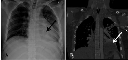
A 25-year-old male belted driver was injured in a car crush in the front collision. He was agitated on the scene and had a prolonged extraction time (45 min). After field intubation the patient arrived to the Trauma Unit under stable hemodynamic and respiratory conditions. He had superficial lacerations of face, left shoulder and both chest and pelvic seatbelt signs. Breath sounds were normal and equal on both sides. A chest radiograph (Figure 1A) and CT scan (Figure 1B) upon admission revealed traumatic rupture of the diaphragm (arrows) on the left side with mild contusion of both lungs. Additionally grade 1 right liver lobe hematoma, grade 1 right kidney laceration and closed non displaced fracture of left rami pubis were found. There were no head and spine injury.

    Figure 1:  An admission chest radiograph (1A) and sagital CT scan (1B)
showed traumatic rupture of the diaphragm (arrows) on the left side with mild
contusion of both lungs. Part of stomach is seen in the left hemithorax.
The patient was transferred to the operating theater 4 hours after arrival and underwent diagnostic laparoscopy. Intraoperative exploration showed that the fundus of the stomach and greater omentum had been transposed into the thorax through a 5 cm tear in the left diaphragm just next to the left diaphragmatic crura underneath the pericardium.
Operative technique
Laparoscopic repair of a traumatic diaphragmatic hernia was performed in a 30° reverse Trendelenburg position. The surgeon stands between the patient’s legs, one assistant to the left and one to the right of the patient.
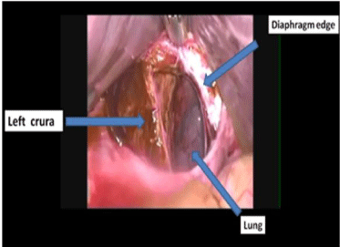
CO2 gas is insufflated using an 11 mm Endopath Xcel port (Ethicon, Endo surgery, Puerto-Rico, USA) inserted through a paraumbilical incision. A 30° angle laparoscope is introduced. The abdominal cavity is explored for free fluids and possible bowel injury. In our case two 12-mm and one 5-mm working ports were placed in a half-circle in the right and left to middle in the upper abdomen so that the stomach could be retracted. Liver retractor (Buchwalter Mediflex) was placed through the additional trocar in the right upper abdomen. After the stomach and greater omentum were repositioned, left pleural cavity was inspected in its entirety (Figure 2).

    Figure 2:  Diaphragmatic tear left to the crura after stomach was reduced
from pleural cavity.
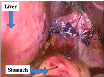
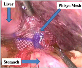
Following complete preparation of the torn rim of the diaphragm, the opposing edges were sutured using nonabsorbable Ethibond 2-0 sutures (Ethicon) tied intracorporeally. An absorbable 5 mm stapler (Secure strap, Ethicon) was used to reinforce the suture line with single staples (Figure 3). Phisyo flexible composite mesh (Ethicone, 15X15 cm) was used to cover the suture line and reinforce the repair (Figure 4). Chest drain was left in the left chest cavity for 48 hours.

    Figure 3:  Suture line after primary repair of diaphragmatic tear with nonabsorbable
sutures.

Figure 4: Composite mesh positioned to reinforce suture line.
Following uncomplicated course, patient was discharged home. No complications are recorded during a year of clinic follow up.
Discussion
TDI is of a great concern for the trauma surgeon. With associated mortality rate more than 20% [1], Plain chest film may raise suspicion for TDI. CT showed a good sensitivity, specificity, negative predictive value and positive predictive value in diagnosing of TDI [2]. It is the diagnostic modality of choice in a resuscitated, stable patient. In present case CT allowed reconstruction in the coronal and sagital plane and gave a definitive diagnosis of diaphragmatic rupture, its location and as a result to carry out treatment plan.
There are several surgical approaches to address TDI. Laparotomy allows exploring the abdominal cavity and assessing the concurrent injuries. It is absolutely preferable when intraabdominal injury cause hemodynamic instability. Minimally Invasive Surgeries (MIS) such as laparoscopic, thoracoscopic or Video Assisted Thoracoscopic Surgery (VATS) approach should also be taken into consideration. VATS has almost 100% specificity and sensitivity in diagnosing TDI in the OR setting, but limited to one hemidiaphragm [3]. The use of therapeutic laparoscopy should be restricted to the treatment of trauma patients with an isolated diaphragmatic injury, because the rate of missing associated abdominal injuries is as high as 41% [4]. In the chronic setting, laparoscopy is a useful tool for both diagnosis and treatment.
Surgeon’s skill and experience should be taken into account when performing a MIS approach to repair a diaphragmatic defect. Exposure is more difficult for posterior diaphragmatic wounds due to patient positioning and organ retraction. We used to position patient with left side up (30 degree). It is also important to insert a chest tube in the side of the diaphragmatic injury to prevent the development of a tension pneumothorax from abdominal insuflation. In our case the diaphragmatic injury located close to the esophageal hiatus was addressed through laparoscopic approach with the same advantages as other surgical procedures on the lower esophagus (for example: Heller myotomy, Nissen fundoplication). Primary suture of diaphragm is a mainstay of surgical treatment. Large and especially chronic defects may require prosthetic patch [5]. We used mesh in our case to reinforce primary repair in hiatal area in order to reduce tension on suture line. In patients with significant tension after primary closure or when the approximated tissue appears weak the use of a prosthetic mesh outweighs the risk of infection and erosion to adjacent organs.
Conclusion
Laparoscopic approach both diagnostic and therapeutic became common in penetrating injury to diaphragm in hemodynamically stable patients. In blunt trauma TDI resulted from high energy impact and associated with serious injuries. CT scan allows recognizing about 80% of diaphragmatic tears. If concomitant injuries can be ruled out, laparoscopy may be considered. Protocols for the selection of these patients are not currently in place and are a promising area of continued research.
References
- Zarour AM, El-Menyar A, Al-Thani H, Scalea TM, Chiu WC. Presentations and outcomes in patients with traumatic diaphragmatic injury: a 15-year experience. J Trauma Acute Care Surg. 2013; 74: 1392-1398.
- Larici AR, Gotway MB, Litt HI, Reddy GP, Webb WR, Gotway CA, et al. Helical CT with sagittal and coronal reconstructions: accuracy for detection of diaphragmatic injury. Am J Roentgenol. 2002; 179: 451-457.
- Dirican A, Yilmaz M, Unal B, Piskin T, Ersan V, Yilmaz S. Acute traumatic diaphragmatic ruptures: A retrospective study of 48 cases. Surg Today. 2011; 41: 1352-1356.
- McCutcheon BL, Chin UY, Hogan GJ, Todd JC, Johnson RB, Grimm CP. Laparoscopic repair of traumatic intrapericardial diaphragmatic hernia. Hernia. 2010; 14: 647-649.
- Hanna WC, Ferri LE. Acute traumatic diaphragmatic injury. Thorac Surg Clin. 2009; 19: 485-489.