Case Report
Austin J Trauma Treat. 2015; 2(1): 1006.
Surgical Management of Traumatic Aorta-Right Ventricular Fistula and Aortic Valve Perforation
Daqing Jiang, Zhi-Wei Zhang*, Xuan Jiang and Qu Chen
Department of Cardiovascular Surgery, The First Hospital of China Medical University, China
*Corresponding author: Zhi-Wei Zhang, Department of Cardiovascular Surgery, The First Hospital of China Medical University, 155 Nanjingbei St, Shenyang 110001, China
Received: November 23, 2015; Accepted: December 17, 2015; Published: December 18, 2015
Abstract
A 22-year-old man had attempted to commit suicide using a knife to penetrate the left anterior chest wall. An emergency operation was performed successfully to repair the penetrating cardiac injury on the Right Ventricular Outflow Tract (RVOT) without cardiopulmonary bypass at local hospital. Four years after the operation, he was found of a continuous murmur over chest wall on a routine check-up. Echocardiography revealed aorto-right ventricular fistula in the sinus of Valsalva with moderate aortic regurgitation. In operation, the perforation of the right coronary cusp and the fistula between aorta and right ventricle were identified. The fistula was closed with a Dacron patch and the aortic valve perforation was repaired with autologous pericardium. Long-term follow-up of penetrating thoracic injuries is important for detecting underlying intra-cardiac lesions.
Keywords: Aortic valve insufficiency; Aorto-right ventricular fistula; Chest stab wound
Case Presentation
Penetrating stab wound to the heart not only penetrate the heart but also damage the intracardiac structure such as valves, intraventricular septum, coronary arteries that would affect long-term outcomes of patients [1]. So far, only a few cases of Aorto-Right Ventricular (Ao-RV) fistula with aortic valve injury have been reported in the English literature [2]. Here, we presented a case with Ao-RV fistula and aortic valve insufficiency 4 years following the initial surgery.
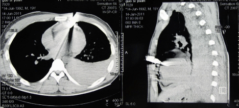
A 22-year-old man had attempted to commit suicide using a knife to penetrate the anterior chest wall 4 years ago. On arrival at the emergency room in a local hospital, he was in shock, with cardiac tamponade and a large left-sided hemothorax (Figure 1). An emergency operation was performed to repair the penetrating cardiac injury. The wound on the surface of the Right Ventricular Outflow Tract (RVOT) was successfully repaired without cardiopulmonary bypass. The postoperative course was uneventful and he was discharged without further checkup or being followed up. Four years after the initial operation, a continuous murmur was heard at the third intercostal space on a routine check-up 2 months before admission. Chest X-ray showed slightly increased pulmonary blood flow and cardiomegaly. ECG showed left ventricular hypertrophy with normal sinus rhythm.

    Figure 1:  A knife penetrating the chest wall with cardiac tamponade and a
large left-sided hemothorax 4 years ago.
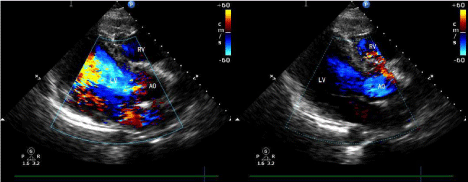
Echocardiography revealed enlarged left ventricule and shunt flow from the right sinus of valsalva to the RVOT with moderate aortic regurgitation (Figure 2). Median sternotomy was performed and the adhesive pericardium was dissected. On inspection of the right ventricular infundibulum, an area of fibrosis with suture was observed 30 mm proximal to the pulmonary valve annulus. Cardiopulmonary bypass was instituted with double venous cannulation under mild hypothermia. After cross clamping, the ascending aorta was opened.

    Figure 2:  Echocardiography revealed moderate aortic regurgitation and
shunt flow from the right sinus of valsalva to RVOT preoperatively.
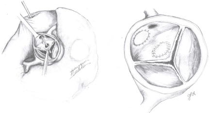
Cardiac arrest was attained by the infusion of cold-blood cardioplegia directly into the coronary ostia. A 6-mm opening was present in the right valsalva sinus, situated between the right coronary ostium and the commissure between the right and left coronary cusps. A 8-mm perforation with fibrosed margins was present in the corresponding area of the right coronary cusp. The RVOT was opened transversely over the area of fibrosis and a 9-mm slit-like opening with fibrosed margins was identified. A vascular clamp could be passed through this opening into the right valsalva sinus .The right ventricular aspect of the fistula was closed with a Dacron patch. The perforation on right aortic cusp was repaired with autologous pericardial patch (Figure 3). After rewarming, the heart resumed sinus rhythm automatically and intraoperative Transesophageal Echocardiography (TEE) showed only trivial aortic regurgitation. The post-operative course was uneventful. Transthoracic Echocardiography (TTE) on day five postoperatively confirmed the absence of left to right shunt and well-functioning aortic valve. The patient was discharged on postoperative day six.

    Figure 3:  The right ventricular aspect of the fistula was closed with a Dacron
patch. The perforation on right aortic cusp was repaired with a autologous
pericardial patch.
Discussion
The time interval between injury and surgical intervention is variable. Some patients require immediate surgical management due to instable hemodynamics, but others may have a delayed clinical presentation and therefore a delayed repair. The interval until definitive repair could be as long as 17 years, as reported by Ehrenstein and colleagues [3]. In our case, the time interval between injury and complete repair was 4 years. All patients with traumatic aorta-right ventricular fistula combined with aortic insufficiency were operated on sooner or later, as reported in the review by Samuels and colleagues [4]. It has been confirmed that a traumatic aorto-right ventricular shunt with aortic insufficiency has a tendency to increase in size with time. Therefore it is advisable that these patients be operated on at an early stage [5]. Although patients with aorto-right ventricular fistula combined with aortic insufficiency after a penetrating trauma may have no cardiac symptoms, they should be thoroughly evaluated, preferably by TEE, and operated on during the same admission. If left untreated, congestive heart failure will invariably develop.
References
- Carter RL, Albert HM, Glass BA. Traumatic ventricular septal defect. Ann Thorac Surg. 1967; 4: 256-259.
- Esfahanizadeh J, Tashnizi MA, Moeinipour A, Shamloo AS. Undetected aorto-RV fistula with aortic valve injury and delayed cardiac tamponade following a Chest Stab Wound: A Case Report. Trauma Mon. 2013; 18: 95-97.
- Ehrenstein FL, Bahler RC, Ankeny J, Schwartz H. Untreated (combined) intracardiac and valvular trauma with long asymptomatic survival. Am Heart J. 1971; 81: 685-687.
- Samuels LE, Kaufman MS, Rodriguez-Vega J, Morris RJ, Brockman SK. Diagnosis and management of traumatic aorto-right ventricular fistulas. Ann Thorac Surg. 1998; 65: 288-292.
- Kaya A, Dekkers P, Loforte A, Jaarsma W, Morshuis WJ. Traumatic aorto-right ventricular fistula with aortic insufficiency. Ann Thorac Surg. 2005; 80: 2362-2364.