
Research Article
Austin J Trauma Treat. 2016; 3(1): 1012.
Prevalence of Chondral Lesion in Knee Arthroscopy
Bikash KC¹*, Lamichhane AP² and Mahara DP²
¹Mechi Zonal Hospital, Nepal
²Department of Orthopaedics, IOM, TU Teaching Hospital, Nepal
*Corresponding author: Bikash KC, Mechi Zonal Hospital, Bhadrapur Jhapa, Nepal
Received: August 01, 2016; Accepted: November 04, 2016; Published: November 10 , 2016
Abstract
Introduction: Chondral lesions are painful and disabling, have a poor capacity for repair, and may predispose patients to early osteoarthritis. Chondral lesions are difficult to diagnose clinically because meniscal injuries also have similar features and clinical signs have low predictive value and specificity. Arthroscopy is the definitive modality of diagnosis. The purpose of this study is to provide a data on prevalence of chondral lesions in patients with symptomatic knees requiring arthroscopy. So that probable outcomes in terms of pain and swelling of the knee following arthroscopy can be explained to the patient.
Method: This was hospital based prospective observational study. There were 75 patients (45male and 30female) of the age group 18-50 years included in this study. Patients having symptomatic knee pain were screened clinically and those fulfilling inclusion criteria were included in study. Patient profile, clinical and arthroscopic findings were recorded in proforma. Chondral lesions were graded according to Outer bridge classification system.
Results: Chondral lesions were found in 49.33% (n=37) of all patients (n=75) with average age of 29 years. Most of the patients were students and average duration of symptoms was 18 months. In these patients, 60% related their current knee problem to a previous trauma. Outer bridge Grade III accounted 46% of all lesions. Of the chondral lesions, 50% were in the medial femoral condyle, 17% in patella, 16% in lateral femoral condyle and 10% in trochlea. Concomitant medial meniscus injury, anterior cruciate ligament injury or both were found in 19%, 13%and 17% of the cases respectively.
Conclusion: Our study supports that articular cartilage defects are common findings in knee arthroscopy. So patients with meniscus like symptoms but not diagnostic clinically or radiologically should always have the suspicions of chondral lesion. However the findings could not be generalized because of relatively small sample size.
Keywords: Chondral lesion; Knee arthroscopy; Osteoarthritis; Meniscus injury
Introduction
The normal function of the knee joint depends on the presence of smooth surface with a low friction index provided by articular cartilage. Articular cartilage is a complex tissue that is able to withstand tremendous forces over many cycles but does not have the ability to heal even after a minor injury [1-6]. Patients with articular cartilage injury usually complains of pain, effusion, and mechanical symptoms. Vulnerability of articular cartilage to various types of lesions may restrict the proper knee function and may lead to osteoarthritis [7]. Chondral lesions are difficult to diagnose; clinical signs like pain, crepitation, effusion, and decrease in movement have a low predictive value and specificity. Patients having chondral lesion may present with meniscus like symptoms. Diagnosis of cartilage lesions can be made by MRI. However, its validity strongly depends on the technique and the radiologists’ personal experience. Arthroscopy has become an accurate method of diagnostic assessment and a surgical technique for therapeutic procedures of the knee joint. It has a diagnostic accuracy of over 90% and is recognized as the gold standard in investigation of the knee [8,9]. The number of patients visiting our institution for symptomatic knee pain requiring arthroscopy is significant. The purpose of this study is to provide a data on incidence of chondral lesions in symptomatic knee requiring arthroscopy. So that probable outcomes in terms of pain and swelling of the knee following arthroscopy can be explained to the patient.
Materials and Methods
The prospective analysis of 75 knee arthroscopies was performed. The data for this study were retrieved from the patients.
Inclusion criteria for study were who have undergone Arthroscopy of the knee for the following reason:
1. History of knee injuries and persistent knee pain,
2. Unexplained knee pain and dysfunction,
3. Loose body sensation,
4. Meniscal tears, and
5. ACL reconstruction
For the age group between 18-50 years.
Exclusion Criteria:
1. History of major knee injury
2. Inflammatory Arthritis
3. Infective arthritis
4. Known Osteochondritis Dessicans
5. Patient before18 and after 50 years of age
The detailed evaluation of the patients with the knee pain was done in Sports OPD. The selected patient data were focused on the onset of symptoms (traumatic, non-traumatic), age, gender, mechanism of injury, sports activity. In cases of repeated arthroscopic procedures, only the first procedure was analysed.
Permission was taken from institutional review board of institute of Medicine prior to starting of the study.
Arthroscopies were performed by two surgeons, specialists in orthopaedics and traumatology, who had practiced the arthroscopic surgery for at least 5 years and had been working together in one centre for more than 10 years. Using the same tools for analysing the chondral pathology (arthroscopic instruments, lesion classification system) they had achieved a high level of agreement regarding the arthroscopic estimation of the cartilage lesion.
The proforma providing information regarding cartilage lesion (grade, location, status of surrounding cartilage), associated articular lesion and the performed procedure was completed after each arthroscopy by the observer. The lesion grade was determined according to the Outer bridge classification [10]. The location of the lesion was documented on the articular surfaces of patella, medial femoral condyle, lateral femoral condyle, trochlea, medial tibial plateau, lateral tibial plateau and the size of the lesion was estimated with the use of a meniscal probe (4 mm). The collected date was entered in SPSS (Statistical Package for Social Sciences) 17.0 program and analysed. Microsoft word and Microsoft Excel 2007 were used for the creation of tables and graphs.
Results
Chondral lesions were found in 37 (49.3%) of 75 arthroscopies. The study group, with diagnosed cartilage lesion, consisted of 45 male and 30 female patients. The average age of patients was 29.41 years and the largest group was patients aged 21–30 years 56% (Table 1). Most of the patients were students (n=27, 36%).
![]()
    Age    Group
    Number
    Percentage


    <20 Years
    7
    9%


    21-30    Years
    42
    56%


    31-40 Years
    14
    19%


    41-50 Years
    12
    16%


    Total
    75
    100%
Table 1: Age distribution of patients.
The analysis of the onset of symptoms revealed that in 60% (n=45) it was a traumatic onset, usually connected with a day living activity and with sports participation. Knee pain (42%) and giving way (25%) were the main complains of the patients. The mean duration of injury was 18.76 months while the longest time being 6 years and shortest 1 month.
The analysis of the localization of the lesion revealed that the medial femoral condyle (50%) and patellar articular surface (17%) were the most frequent localizations of the cartilage lesions, while medial tibial plateau (7%) was the least frequent one (Figure 1).
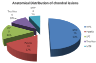
Figure 1: Anatomical distribution of chondral lesions.
Grade III according to Outer bridge classification, evaluated in arthroscopy, was the most frequent grade of the cartilage lesion (46%). Grade III and IV lesions were documented in 66%. In the group of patients with grade III and IV lesions the predominant age was less than 40 years. Grade III and IV lesions were predominant within the medial femoral condyle and patella (Figure 2).
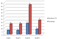
Figure 2: Grading of chondral lesion.
During arthroscopy, chondral lesions were associated with concomitant articular findings like meniscal injury or ligamentous injury. Out of 37 chondral lesion positive patients 7(19%) had medial meniscal injury and 5(13%) had torn Anterior Cruciate Ligament (ACL) and 6(17%) had both menisci and anterior cruciate ligament tear. Distribution of other concomitant arthroscopic findings is shown in Table 2.
![]()
    Arthroscopic Findings
    No. in chondral lesion (n=37)
    Percentage


    MMT
    7
    19%


    MMT+LMT
    3
    8%


    ACL+MMT
    7
    19%


    ACL+MMT+LMT
    6
    17%


    ACL    Tear
    5
    13%


    ACL+LMT
    1
    3%


    LMT
    3
    8%


    Chondral    lesion only
    5
    13%


    Total
    37
    100%


    MMT: Medial meniscus Tear; LMT: Lateral Meniscus Tear;    ACL: Anterior Cruciate Ligament
Table 2: Associated arthroscopic findings.
Discussion
Articular cartilage lesions are a common pathology of the knee joint. Since the times of Hippocrates it has been observed that cartilage once damaged never heals. So far the natural history of cartilage lesions remains unpredictable and not well understood. Arthroscopy has evolved to become an accurate method of diagnostic assessment and a surgical technique for therapeutic procedures of the knee joint. It has a diagnostic accuracy of over 90% and is recognized as the gold standard in investigation of the knee [8,9]. This study was carried out to provide data on the incidence of cartilage lesions in knee arthroscopy and classify them according to Outer bridge classification system [10] including their anatomical location.
In this study, age group of 18-50 years patients were taken and the mean age of the patients was 29.41 years with largest group were patient aged 21-30 yrs 56%(n=42). The lower age was chosen in such a way that after 18 years, the skeletal maturity and chondral calcification is complete. In skeletally immature and young adults osteochondral fractures are more common than chondral lesions [11,12]. Similarly, after 50 years, 80% develop features of degenerative changes [13,14]. This age group (21-30) is highly engaged in athletic activities and other physical activities and hence more likely to get knee injuries.
Male patients were common than female patients representing 60% (n=45) and 40% (n=30) respectively. Our finding is similar to findings of David Figueroa et al. [15], Hjelle K et al. [16], Widuchowski J et al. [17], Curl WW et al. [18]. Male predominance in knee injuries may be due to the fact that males are more involved in sports as well as in outdoor activities and road traffic accidents whereas females remain busy in indoor activities.
In this study analysis of onset of symptoms showed that 45(60%) had traumatic injury and 30 (40%) had no history of trauma. Sports injury was common 25(56%) among the traumatic onset group. Similarly, Widuchowski J et al. [17] analysied of the onset of symptoms revealed that in 58% patients had a traumatic non-contact onset, usually connected with a day living activity (45%) and with sports participation in 46%. Hjelle K et al. [16] stated 61% of patients sustaining trauma of the knee leading to present complains. The commonest mode of injury in this study was sports like high-jump, long-jump, volleyball and football followed by blunt trauma and road traffic accident. Twisting injuries with or without carrying weight was a significant mode of injury.
In this study Knee Pain, giving way and locking were the common presenting complains of the patients each 42%, 25% and 18% respectively. There may be more than one complains because of relatively late presentation of patients for arthroscopy (18 months) and there were associated intra-articular injuries like ACL tear and Meniscal tear.
Mean time period form onset of symptoms and current arthroscopy in our study was 18.76 months (range 1-72 months). Such a long duration of injury might be due to many factors such as lack of expertise to diagnose such injuries in the peripheral areas in our country, lack of proper referral system, lack of awareness or ignorance of the injury by patients till it becomes disabling.
In this study, Chondral lesions were found in 49.33% (37) of 75 arthroscopies. In study of David Figueroa et al. [15] incidence of chondral lesion was 41.16% (82 in 190 cases). In a Analysis of articular cartilage lesions in 5114 knee arthroscopies by Widuchowski W, Kusz D, et al. [17] chondral lesions were found in 57.3% Hjelle et al. [16] evaluated 1,000 knee arthroscopies and identified chondral lesions in 61% of the patients. Curl W et al. [18] with 35,516 arthroscopies had 63% incidence of chondral lesion and Aroen A et al. [7] found 66% in 993 knee arthroscopies. Overall the incidence of chondral lesion ranged from 40-65% in most of the studies which is similar to this study.
In this study most common location of chondral lesion was medial femoral condyle 29(50%), followed by patella 10(17%) and lateral femoral condyle 9(16%). Medial femoral condyle and patella were common location of chondral lesions in other studies also. Hjelle et al. [16] found 58% of chondral lesion in the medial femoral condyle, 11% in patella, 11% in lateral tibia, 9% in lateral femoral condyle, and5% in medial tibia. Widuchowski, J et al. [17] revealed that the patellar articular surface (36%) and the medial femoral condyle (34%) were the most frequent localizations of the cartilage lesions, while medial tibial plateau (6%) was the least frequent one. Similarly, Aroen A et al. [7] identified 43% of lesions in medial femoral condyle followed by 23% in patella. Curl W et al. [18] also stated medial femoral condyle (32%) and patella (21%) as common locations. David Figueroa et al. [15] showed a predilection for the medial femoral condyle (32.2%), the medial articular surface of the patellae (22.6%), and the lateral femoral condyle (14.8%).
In this study Grade III according to Outer bridge classification was the most frequent grade of the cartilage lesion 46% (n=37) followed by Grade IV 22%. Grade I and II lesions were documented in 16% each. This finding was similar to findings of Hjelle et al. [16] (55% grade III and 5% grade IV lesions 14% grade I, 26% were grade II) and Curl WW et al. [18] (9.7% grade I; 28.1% grade II; 41.0% grade III; and 19.2% grade IV lesions respectively).
In this study concomitant medial meniscus injury was found in 19% patients followed by both meniscus and ACL tear in 17% patients and 13% had only ACL tear. In the study of Hjelle K et al. [16], concomitant meniscal or anterior cruciate ligament injury was found in 42% and 26% respectively. Concomitant injury of both meniscus and anterior cruciate ligament injury was found in 12%. Similarly Widuchowski, J et al. [17] found the most common associated lesions were the medial meniscus tear (37%) and the injury of the anterior crucial ligament (36%). In another study by Hjelle K et al. [16] meniscus lesions were found in 57% of the patients, Anterior Cruciate Ligament (ACL) injury in 17%. In most of studies medial meniscus tear and ACL tear are common associated findings with chondral lesion which is similar to our study.
In this study of the 41 patients who had ACL insufficiency, concomitant chondral lesion were observed in 19 patients (46.34%) and 13(31.70%) were Grade III, IV. Medial meniscus tear was present in 18(43.9%), both menisci tear in 5(12.19%) patients and Lateral meniscus tear in 3(7.31%). In 1993 Spindler et al. [19] prospectively studied 54 consecutive patients who underwent arthroscopic ACL reconstruction. Articular cartilage defects were recorded at the time of surgery in 46% of knees which is similar to this study.
Conclusion
In conclusion, chondral lesions are common findings in knee arthroscopy, present in almost half (49.33%) of the arthroscopies performed (n=75) in this study. Most of lesions were of Outer bridge Grade III (46%), and a significant proportion of chondral lesions were associated with other intra articular disorders like ACL tear and medial meniscus tear. The lesions were found most often in the medial femoral condyle (50%), and more often in the patients under 40 years of age. So patients with meniscus like symptoms but not diagnostic clinically or radiologically should always have the suspicions of chondral lesion. However the findings could not be generalized because of relatively small sample size.
References
- Buckwalter JA, Mankin HJ. Articular cartilage: Tissue design and chondrocytematrix interactions. Instr Course Lect. 1998; 47: 477-486.
- Buckwalter JA, Mankin HJ. Articular cartilage: Degeneration and osteoarthritis, repair, regeneration, and transplantation. Instr Course Lect. 1998; 47: 487-504.
- Lefkoe TP, Trafton PG, Ehrlich MG, Walsh WR, Dennehy DT, Barrach HJ, et al. An experimental model of femoral condylar defect leading to osteoarthrosis. J Orthop Trauma. 1993; 7: 458-467.
- Bauer M, Jackson RW. Chondral lesions of the femoral condyles: A system of arthroscopic classi?cation. Arthroscopy. 1988; 4: 97-102.
- Caplan AI, Elyaderani M, Mochizuki Y, Wakitani S, Goldberg VM. Principles of cartilage repair and regeneration. Clin Orthop. 1997; 342: 254-269.
- Twyman RS, Desai K, Aichroth PM. Osteochondritis dissecans of the knee. A long-term study. J Bone Joint Surg Br. 1991; 73: 461-464.
- Aroen A, Deryk G, Jones DG, Fu FH. Arthroscopic diagnosis and treatment of cartilage injuries. Sports Med Arthrosc Rev. 1998; 6: 31-40.
- Tait GR, Maginn P, Macey AC, Beverland DE, Mollan RAB. Unnecessary arthroscopies. Injury. 1992; 23: 555-556.
- Fife RS, Brandt KD, Braunstein EM, et al. Relationship between arthroscopic evidence of cartilage damage and radiographic evidence of joint space narrowing in early osteoarthritis of the knee. Arthritis Rheum. 1991; 34: 377- 382.
- Outerbridge RE. The etiology of chondromalacia patellae. J Bone Jt Surg. 1961; 43B: 752-757.
- Buckwalter JA, Woo SL-Y, Goldberg VM, Hadley EC, Booth F, Oegma TR, et al. Soft tissue aging and musculoskletal function. J Bone Joint Surg Am. 1993; 75A: 1533-1548.
- Buckwalter JA, Lane NE. Aging, sports and osteoarthritis. Sports Med Arth Rev. 1996; 4: 276-287.
- Johnson LL. Arthroscopic abrasion arthroplasty. McGinty JB, editor. In: Operative Arthroscopy. NewYork, NY: Raven Press. 1991; 341-360.
- Meunier A. Osteoarthritis after surgical or conservative treatment of the acutely torn anterior cruciate ligament — a randomised study with 15 years follow-up. Swedish Orthopaedic Society. Acta Orthopaedica Scand. 1999.
- Figueroa D, Calvo R, Vaisman A. Knee Chondral Lesions: Incidence and Correlation between Arthroscopic and Magnetic Resonance Findings. Arthroscopy: The Journal of Arthroscopic and Related Surgery. 2007; 23: 312-315.
- Hjelle K, Solheim E, Strand T, Muri R, Brittberg M. Articular cartilage defects in 1,000 knee arthroscopies. Arthroscopy. 2002; 18: 730-734.
- Widuchowski W, Kusz D, Widuchowski J, Faltus R, Szyluk K. Analysis of articular cartilage lesions in 5114 knee arthroscopies. 2006.
- Walton WC, Jonathan K, Stanly G, Julia R, Beth PS, Gary GP. Cartilage injuries: A review of 31,516 knee arthroscopies. J Arthroscopy. 1997; 13: 456-460.
- Spindler KP, Schils JP, Bergfeld JA, et al. Prospective study of osseous, articular, and meniscal lesions in recent anterior cruciate ligament tears by magnetic resonance imaging and arthroscopy Am J Sports Med. 1993; 21: 551-557.