Clinical Image
Austin J Trauma Treat. 2022; 7(1): 1017.
Isolated Greater Trochanteric Fracture in Elderly
Zaizi A*, ElHassak S and Boussouga M
Department of Orthopaedic Surgery & Traumatology II, Mohamed V Military Hospital, Faculty of Medicine and Pharmacy, Mohamed V University, Morocco
*Corresponding author: Abderrahim Zaizi, Doctor at Department of Orthopaedic Surgery & Traumatology II, Mohamed V Military Hospital, Faculty of Medicine and Pharmacy, Mohamed V University, Rabat 10100, Morocco
Received: October 10, 2022; Accepted: November 07, 2021; Published: November 14, 2022
Clinical Image
Isolated greater trochanter fractures are unusual presentation following direct hiptrauma, most often low energy in elderly patient [1]. Traditional these injuries are managed non-operatively by bed rest, taping or hip spica casting. However, available new imaging such as CT scans or MRI imaging should be indicated to look for the extent of the lesion, leading to choose adequate and safe treatment. Schultz states that intertrochanteric fractures that do not cross the midline on MRI may be treated conservatively [2].
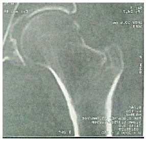
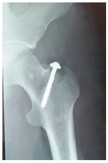
We present a case of isolated greater trochanter fracture of left hip in a 70 years-old patient, CT scan have showed fracture line extending from the greater trochanter without crossing the midline (Figure 1). Thus, patient underwent operative treatment through open reduction and internal fixation by spongious screw and washer (Figure 2).

Figure 1: CT-scan showed fracture line extension from the greater trochanter.

Figure 2: Postoperative X rays.
Conflict of Interest
The authors declare that they have no competing interests.
References
- Prommik P, Tootsi K, Veske K, Strauss E, Saluse T, et al. Isolated greater trochanter fracture may impose a comparable risk on older patients’ survival as a conventional hip fracture: a population-wide cohort study. BMC Musculoskelet Disord. 2022; 23: 394.
- LaLonde B, Fenton P, Campbell A, Wilson P, Yen D. Immediate weightbearing in suspected isolated greater trochanter fractures as delineated on MRI. Iowa Orthop J. 2010; 30: 201-4.