
Case Report
Austin J Trop Med & Hyg. 2015;1(2): 1006.
An Unusual Presentation of Nocardiosis - A Report of Two Cases
Subhasish Ghosh1, Dipankar Pal2*, Shekhar Pal3, Jayanta Roy4 and Arpita Bhakta5
1Consultant Pulmonologist, Apollo and AMRI Hospitals, Kolkata, India
2RMO cum Clinical Tutor, Dept.of Tropical Medicine, School of Tropical Medicine, Kolkata, India
3Assistant Professor, Department of Tropical Medicine, School of Tropical Medicine, Kolkata, India
4Consultant Neurologist, Department of Neurology, Apollo Hospitals, Kolkata, India
5Head of the Dept.of Microbiology, AMRI Hospitals, Kolkata, India
*Corresponding author: Dipankar Pal, RMO cum Clinical Tutor, School of Tropical Medicine, India
Received: December 18, 2014; Accepted: February 17, 2014; Published: February 19, 2015
Abstract
Nocardiosis is an opportunistic infection more common in immune compromised hosts. Disseminated nocardiosis has a poor outcome. We report a case of disseminated nocardiosis with nocardaemia (case-1) which is an extremely rare finding even in immune compromised subjects. In case-2 we found pulmonary abscess caused by nocardia in a patient of sarcoidosis on steroids. Vascular thrombosis complicating nocardiosis is not recognized. We report two cases of nocardiosis with arterial thrombosis.
Keywords: Immunocompromised state; Disseminated nocardiosis; Cerebral abscess; Pulmonary abscess; Vascular thrombosis
Introduction
Nocardia species are saprophytic aerobic actinomycetes and are common worldwide in soil causing decay of organic matter. It is an opportunistic pathogen causing significant morbidity and mortality in human beings. It predominantly affects lung with pre-existing structural defects and also with co-existing mycobacterial infection. Disseminated nocardiosis occurs through haematogenous spread to distant organs including brain (commonest), bone, soft-tissues and kidney; whereas peritoneum and heart valves only rarely affected. Isolating nocardia in blood culture (nocardaemia) is extremely rare [1]. Endovascular foreign body [2] e.g. prosthetic heart valve is a unique risk factor for nocardaemia but our patient (case I) did not posses any such foreign body. Nocardia bacteraemia is also associated with simultaneous infection with other bacterial pathogens, especially Gram negative organisms in 30% [2]. First patient had concomitant Klebsiella infection [Extended-Spectrum Beta-Lactamase (ESBL) producer] in lung. Surprisingly in both the cases vascular thrombosis complicated the picture-thrombosis in pulmonary trunk was in case- 1 and lacunar infarcts of brain found in case-2. Nocardiosis and vascular thrombosis may be causally related.
Case Report
Case I
A 52 yr old gentleman presented to our clinic with fever and dry cough for 15 days. He also complained of chest tightness and exertional breathlessness for the same duration. His fever was low grade, intermittent in nature and subsided only on taking antipyretics. It was associated with cough without expectoration, hemoptysis or chest pain.
He had a significant past history. Ten years ago he was treated for pulmonary tuberculosis.
He presented with foot drop in another institution four months before where he was diagnosed as having mononeuritis multiplex based on NCV/EMG study showing bilateral peroneal neuropathy. His connective tissue panel (Anti-nuclear antibody etc.) and vasculitis screening (ANCA) were all negative at that time. He was started on oral cyclophosphomide (50 mg/day) and prednisolone (40mg/ day) for mononeuritis multiplex in that institute with significant improvement of his muscle strength and became ambulatory. He was also on oral anticoagulant therapy following a massive pulmonary thromboembolism which he suffered few weeks after starting immunosuppressive therapy (Figure 1). At the time of admission, he was receiving prednisolone-40mg/day, cyclophosphomide-50mg/ day, oral anticoagulant and calcium supplement.
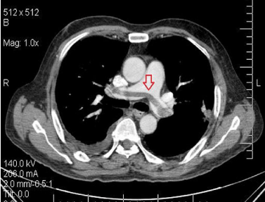
Figure 1: CT Pulmonary Angiography showing saddle embolus.
Examination of the respiratory system revealed tachypnoea and coarse crackles in right infrascapular area. Neurological examination revealed wasting of proximal and distal muscles of both lower limbs with muscle power of 4/5 in all groups. Deep tendon reflexes were preserved except left knee jerk. Sensory system, cranial nerves and cerebellar functions were all normal.
Total leucocyte count was 15,500/cu mm with neutrophilia, procalcitonin-0.14 (normal < 0.05), INR-5.27. Platelet count-3.80lac/ cu mm. Blood culture was sent. Chest x-ray showed nodular opacity in right lower lobe with pleural reaction (Figure 2). CT thorax with CT pulmonary angiography revealed nodular consolidation involving anterior and lateral segment of right lower lobe with area of necrosis abutting pleura with adjoining pleural reaction. No pulmonary thrombosis or embolism was evident (Figure 3) this time Fibre Optic Bronchoscopy (FOB) and Bronchoalveolar Lavage (BAL) with washing from right basal and apical segment of right lower lobe was done and sent for Gram stain , AFB stain, culture, fungal stain and modified AFB and silver stain.
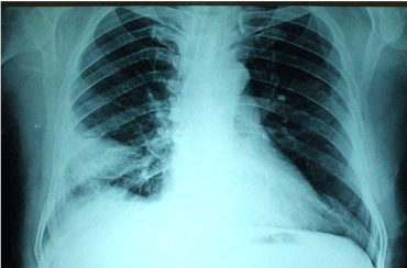
Figure 2: CXR showed nodular opacity in right lower lobe with pleural reaction.
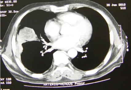
Figure 3: CT Scan thorax showing nodular consolidation involving right lower lobe.
Gram positive branching, fine, delicate, filamentous organisms positive on modified AFB stain (using 1% sulphuric acid as decolourising agent) (Figure 4) [3] was detected on BAL along with growth of Klebsiella aerogenes [ESBL producer] and Nocardia asteroides on culture. Blood culture also grew Nocardia asteroides (growth time 59 hrs) [1]. The colonies of Nocardia grown from both BAL and blood were adherent, dry, wrinkled, chalky white on sheep blood agar .The isolate was identified as Nocardia asteroides by Vitek 2 Compact system (Biomerieux).Isolate was sensitive to ceftriaxone, amikacin, imipenem, co-amoxiclav and trimethoprim-sulfamethoxazole. Trimethoprim-sulfamethoxazole [TMP 640 mg/day in divided doses] and imipenem [4] were started, cyclophosphamide was stopped after discussion with neurologist. A tapering regimen of steroid was initiated. He started to show positive response to above treatment of pulmonary nocardiosis [5] with subsidence of fever. However, on the third day of treatment he complained of giddiness and soon had a brief generalized tonic clonic seizure. CT brain showed a right temporal hypodensity. MRI revealed multiple cerebral abscesses with ring enhancement and perilesional edema (Figure 5) [6]. We started meropenem in place of imipenem and phenytoin was added to control seizure. He remained stable on this combination. We discharged him after a week on above medications and readmitted 2 weeks later for craniotomy and drainage of cerebral abscess. The cerebral abscess material also grew Nocardia asteroides on culture Co-trimoxazole and meropenem were continued for 4 weeks in view of disseminated disease and he was discharged home on oral co-trimoxazole, co-amoxiclav, prednisolone - 5mg daily and oral anti-coagulant with monthly follow-up advice (Table 1). He remained a febrile without neurological sequel, with chest x-ray showing excellent resolution (Figure 6).
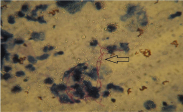
Figure 4: Modified AFB stain showing branched filamentaous organisms.
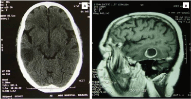
Figure 5: CT Brain showing right temporal hypodense lesion. (B) MRI picture of cerebral abscess.
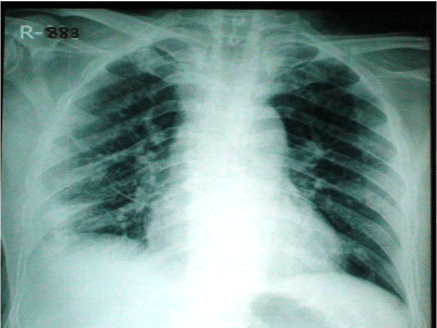
Figure 6: Chest X-ray post discharge showing significant resolusion.
    ANTIBIOTICS
    I
    ANTIBIOTICS
    I


    Penicillin G

    Combinations


    Amoxycillin
    S
    Amoxy-clav
    S


    Pipericillintazobactum
    S


    Vanconnycin
    S
    Aminoglycosides


    Clindamycin
    S
    Gentamycin
    S


    Tobramycin
    S


    Fluroquinolones

    Netilmycn
    S


    Noriloxacin

    Amikacin
    S


    Ciprofloxacin
    S


    Ofloxacin
    S
    Others


    Levofloxacin
    S
    Cotrimoxazole
    S


    Gatifloxacin
    S


    Carbapenems


    Imipenem
    S


    S: Sensitive, R:    Resistant, I: Intermediate
Table 1: Antibiogram of (Case 1).
Case II
A 53 yr old lady was admitted in our institution with a febrile illness for 2 weeks associated with right sided pleuritic chest pain and easy fatiguability . Ten months ago she first presented to a local doctor with persistent cough. A CT thorax at that time showed nodular opacities with some of the nodules along the bronchovascular bundles and consolidation of apical segment of left lower lobe with ground glass opacity. There was widespread mediastinal lymphadenopathy involving right paratracheal, pretracheal, precarinal, AP window and subcarinal stations (Figures 7 & 8). As she refused bronchoscopy, prednisolone (60mg/d) [7] was started with a provisional diagnosis of sarcoidosis. Her cough got better and chest x-ray (Figure 9) showed significant resolution, especially of left lung opacities. There was no history suggestive of pulmonary tuberculosis/connective tissue disease/ischaemic heart disease or asthma. She was a nonsmoker and non-alcoholic but she became diabetic, likely steroid induced.
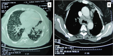
Figure 7: CT Thorax showing (A)nodular lesions in peri lymphatic distribution with ground glass opacity. (B) Mediastinal lymphadinopathy.
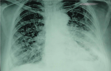
Figure 8: Chest X-ray showing nodular opacities.
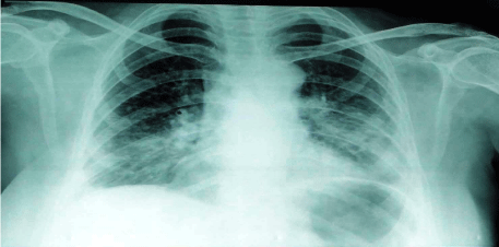
Figure 9: Chest X-ray showing improvement following steroids.
On clinical examination, she had cushingoid facies with oral thrush, was febrile and appeared toxic. There was no palpable peripheral lympadenopathy, engorged jugular veins, pedal edema. Examination of the respiratory system revealed diminished vesicular breath sound with coarse crepitations at places of right infrascapular and infraaxillary area with an evanescent rub. Examination of all other systems was normal. She was on 30 mg/day of prednisolone [7] and chest x-ray revealed dense nodular shadows in right lower zone (Figure 10) [6,9] during admission. Hemoglobin-12.1gm%,total leucocyte count-8100(Neutrophil-87%), platelets-6.87lac/ul, ESR- 103, CRP> 250, Procalcitonin-0.29 (normal<0.05), SGOT/SGPTnormal, GGT-319, Alkaline-phosphatase-222, urea, creatinine, electrolytes-normal. Blood culture was sent, Sputum was sent for gram stain, AFB stain and culture.
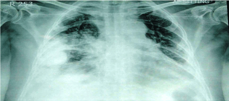
Figure 10: Chest X-ray showing dence nodular opacities involving right lower lobe.
Management was focused in the line of pneumonia in an immune compromised host (steroid induced). A tapering regime of prednisolone was initiated. Broad spectrum antibiotic cover was commenced with ertapenem, linezolid and moxifloxacin. CT scan thorax showed right middle and lower lobe necrotizing pneumonia with right sided multi loculated pleural effusion (Figure 11) [6,9]. The opacity at apical segment of left lower lobe had regressed considerably with sparse nodules. Blood culture was unyielding. Sputum examination however revealed plenty of gram positive, beaded, branched filamentous organisms suggestive of nocardia species which was also positive on modified acid fast stain. Ertapenem was changed to meropenem [4], linezolid was continued and co-trimoxazole was started (TMP 15mg/kg/day) in place of moxifloxacin.
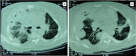
Figure 11: CT Scan showing (A)Right middle lobe consolidation with air bronchogram. (B) showing right sided multiloculated pleural effusion.
After being put on this regimen she became a febrile in 48 hours and continued to show good clinical response. CT scan brain did not show any abscess. MRI brain however showed tiny foci of lacunar infarcts in temoro-parietal (bilateral) and left occipital regions. She remained a febrile with the above regime. In the meantime, sputum culture yielded Nocardia asteroides. Meropenem was stopped after 14 days and co-amoxiclav was added based on the sensitivity pattern of the organism in sputum culture. Linezolid was also stopped as there was no evidence of disseminated disease.
Patient was discharged after 2 weeks on oral cotrimoxazole and coamoxiclav (Table 2). CRP was 35 on discharge. She was also on maintenance dose of steroid (5 mg in morning and 2.5 mg in evening) and clopidogrel. She continued to do well without drug related adverse events noted so far. Chest X-ray one month post-discharge showed significant improvement (Figure 12).
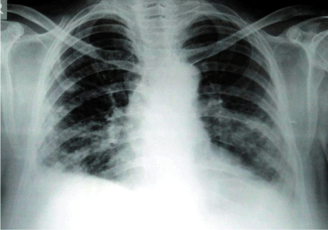
Figure 12: Chest X-ray showing significant improvement with treatment.
SPECIMEN: SPUTUM
METHOD: Aerobic bacterial culture
REPORT : Culture shows growth of Nocardia asteroides
Colony count :104cfu/m1
ANTIBIOGRAM (Disc Diffusion method)
    ANTIBIOTICS
    I
    II
    ANTIBIOTICS
    I
    II


    Penicillins


    Combinations


    Amoxycillin
    S

    Amoxy-clav
    S


    Piperacillin


    Ampicillin-sulbactam


    Ticarcillin


    Piperacillin-tazobacta m


    Ticarcillin-clavulunic acid


    Cephalosporins


    Cefoperazone-sulbactam


    Cefuroxime


    Cefdinir


    Aminoglycosides


    Cefixime


    Gentamycin


    Ceftriaxone
    S

    Tobramycin


    Cefotaxime


    Netilmycn


    Ceftazidime


    Amikacin
    S


    Cefepime


    Fluroquinolones


    Cefamycin


    Norfioxacin


    Cefoxitin


    Ciprofloxacin
    S


    Ofloxacin


    Monobactam


    Levofloxacin


    Aztreonam


    Gatifloxacin


    Carbapenems


    Others


    Imipenem
    S

    Nitrofurantoin


    Meropenem


    Chloramphenicol


    Ertapenem


    Cotrimoxazole
    S


    Erythromycin
    S


    Linezolid
    S


    Tetracycline


    Polymyxin    B


    S: Sensitive, R:    Resistant, I: Intermediate
Table 2:  Antibiogram of (Case 2).

SPECIMEN: SPUTUM

METHOD: Aerobic bacterial culture

REPORT : Culture shows growth of Nocardia asteroides

Colony count :104cfu/m1

ANTIBIOGRAM (Disc Diffusion method)
Discussion
Nocardiosis remains an elusive diagnosis despite its discovery by Edmond Nocard in 1888. There are more than 50 species of nocardia with at least 30 species recognized as pathogenic to human. Fragmented bacterial mycelia enter lungs via aerosol route. Although pulmonary nocardiosis is a common presentation [2/3rds of cases] [5], human to human transmission is not documented. It is a disease unique to immunosuppressed individuals. But 30% of infections are also seen in immune competent hosts [6], Chronic Obstructive Pulmonary Disease (COPD) is a risk factor among such groups [7]. Glucocorticoid therapy and malignancy and its treatment are common risk factors [7]. Pulmonary nocardiosis presents as necrotizing pneumonia with or without cavitation. They may also present as a slowly enlarging pulmonary nodule with or without empyema, confusing with malignancy [6,9]. Subacute onset of fever, cough and constitutional symptoms makes pulmonary tuberculosis a common misdiagnosis [10]. The median time to diagnosis is therefore long (42days to 12 months). Its special staining characteristics [modified Kinyuon procedure] and fastidious nature of growth adds to this problem [7]. Extra-pulmonary nocardiosis occurs in half of all cases of pulmonary disease. Because of the long median time to diagnosis and its neurotropism Central Nervous System (CNS) nocardiosis is a common phenomenon [3,6]. CNS nocardiosis usually present as single or multiple abscesses [6]. Dissemination to the brain heralds a poor prognosis with mortality in some studies reported close to 100% [7]. TMP-SMX (TMP15mg/kg) is the first line of treatment because of its excellent penetration and wide susceptibility. Combination treatment should be prescribed in disseminated disease and in immune compromised hosts [10]. Severe disease is to be treated with parenteral therapy with at least two drugs initially (induction phase).Amikacin, imipenem and 2nd/3rd generation cephalosporins, co-amoxiclav are suitable candidates [4].
The duration of the induction therapy is six weeks which is followed by oral combination of two drugs for total period of twelve months [10]. TMP-SMX (TMP 10mg/kg) and co-amoxiclav are good choices here [8]. Some recommend parenteral therapy all through for CNS infection. Prolonged treatment with combination drugs are advised because the two characteristics of nocardia infection are a) wide dissemination and b)tendency to relapse inspite of appropriate treatment.
Although our selection of antibiotics was purely empiric initially, we saw significant clinical response prompting us to continue with this regime. However routine susceptibility tests were available soon (Tables 1 & 2) and we switched to a targeted regime. Drug susceptibility test is strongly recommended in all cases because of prolonged treatment and variable susceptibility among different species.
Conclusion
We describe two cases of severe nocardiosis presenting as necrotizing pneumonia with pleural reaction in patients on immunosuppressive drugs. Our first case is disseminated nocardiosis with positive blood culture (nocardaemia), an extremely rare finding. The finding of pulmonary nocardiosis in an immune compromised individual should prompt us to get brain imaging studies done before commencing an effective anti-nocardial agent like imipenem which has otherwise excellent activity against nocardia. Brain imaging detected multiple right temporal abscesses in our first case. In the second one, we did not find any abscess in brain but there were lacunar infarct.
Literature does not provide information on whether nocardiosis predisposes individuals to vascular thrombosis. An incidental finding in both of our cases was vascular thrombosis (pulmonary thromboembolism in Case I and lacunar infarcts brain in Case II) which can be explained by heightened platelet aggregation probably secondary to thrombocytosis.
Three unique features drew our attention: a) occurrence of nocardaemia without endovascular prosthesis, b) vascular thrombosis complicating nocardiosis, c) learning the lesson that all cases of pulmonary nocardiosis should be evaluated thoroughly with brain imaging to exclude central nervous system involvement as disseminated nocardiosis can be a silent killer.
References
- Ming-Huang Tuo, Ying-Huang Tsai, Hsiang-Kuang Tseng, Wei-sheng wang. Clinical experiences of pulmonary and bloodstream nocardiosis in two tertiary care hospitals in northern Taiwan. 2000-2004.
- Nocardia bacteremia: report of 4 cases & review of literature. Medicine (Bultimore). 1998; 77: 255-267.
- McNeil MM, Brown JM. The medically important aerobic Actinomycetes: Epidemiology and microbiology. Clin Microbiol Rev. 1994; 7: 357-417.
- Lo W, Rolston KV. Use of Imipenem in treatment of Pulmonary nocardiosis. Chest. 1993: 103; 951-952.
- Lederman ER, Crum NF. A case series and focused review of nocardiosis: clinical and microbiologic aspects. Medicine (Baltimore). 2004; 83: 300-313.
- Beaman BL, Beaman L. Nocardia species: Host-parasite relationships. Clin Microbiol Rev. 1994; 7: 213-264.
- Martinez Tomas R, Menendez Villanueva R, Reyes Calzada S, Santos Durantez M, Vallés Tarazona JM, Modesto Alapont M, et al. Pulmonary nocardiosis: risk factors and outcomes. Respirology. 2007; 12: 394-400.
- Sorrell TC, Mitchell DH, Iredell JR. Nocardia species. Mandell GL, Bennett JE, Dolin R, editors. 6th edn. In: Principles and Practice of Infectious Diseases. Elsevier, Philadelphia. 2005; 916.
- Hwang JH, Koh WJ, Suh GY, Chung MP, Kim H, Kwon OJ, et al. Pulmonary nocardiosis with multiple cavitary nodules in a HIV-negative immunocompromised patient. Intern Med. 2004; 43: 852-854.
- Lerner PI. Nocardiosis. Clin Infect Dis. 1996; 22: 891-903.