Case Report
Austin J Urol. 2014;1(2): 2.
Case Report: Acquired Giant Hydronephrosis Presenting as an Abdominal Mass
Moein MR*
Department of Urology, Shahid Sadoughi Medical University, Iran
*Corresponding author: Moein MR, Department of Urology, Rahnemoon hospital, Shahid Sadoughi Medical University, Yazd-Iran
Received: May 16, 2014; Accepted: August 19, 2014; Published: August 22, 2014
Abdominal mass can be a presentation of a variety of pathologic process. One of the most important causes of it, especially in young children, is hydronephrosis. Hydronephrosis is the dilation of the renal pelvis or calyces. It may be associated with obstruction, but may be present in the absence of obstruction. Among the most common cause of hydronephrosis is congenital anomaly of ureteropelvic junction of kidneys. But acquired hydronephrosis especially in adults also may rarely present as an abdominal mass. As we know obstruction of normal urine outflow results in biochemical, immunologic, hemodynamic, and functional changes of kidneys. So, proper diagnosis and management of this pathologic condition is of at most importance to save renal function.
Case Presentation
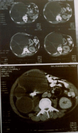
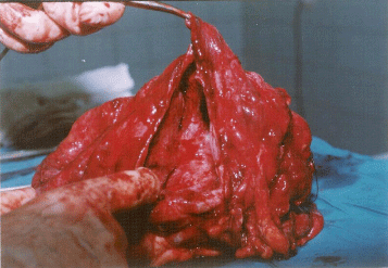
A 47 years old man referred to our urologic clinic due to swelling of right side of abdomen and flank for 6 months. The mass had been gradually larger during this period, but it was not painful until late in the course of illness when it caused patients feel heaviness in flank and abdomen. Patient had no complaints of dysuria, discoloration of urine or any other irritative urinary symptoms. There was negative history of nausea, vomiting, constipation, fever, weight loss, and anorexia. Also he had no history of stone disease and, or any other urologic problem. In physical examination there was no positive finding except a huge soft mass with approximate size of 10×8cm palpated in right sub costal margin and extending laterally to right flank and inferiorly to iliac crest and medially to mid abdomen. The mass was soft with smooth borders and was not tender in palpation. Laboratory data were as follow: Hb: 11.3g/dl, Hct: 35.6, WBC: 6100 BUN: 27mg/dl, Creatinine: 1.0mg/dl. Urinanalysis showed the followings: blood: 1+, WBC: 7-8, RBC: 3-4 and urine culture was negative also. Sonography showed severe right hydronephrosis with pressure effect on abdominal viscera but there was no any intra-abdominal lesion. CT scan with contrast showed severe right hydronephrosis with non-functioning right kidney and a large stone impacted in the first part of the ureter (Figure 1). Isotope scan with TC99-DMSA confirmed right non-functioning kidney. So, patient scheduled for exploration of right kidney and possible right nephrectomy. Through right subcostal incision in right flank position, peritoneum was pushed medially. There was a very huge mass that was filled retroperitoneum completely and shifted abdominal viscera to left side of abdomen. Kidney was punctured and about 3350 cc of clear fluid was evacuated from the kidney after that continuation of the procedure became feasible and right nephrectomy was done. The kidney was very large and thickened even after the evacuation of its fluid as shown (Figure 2). There was also a large impacted stone in the ureteropelvic junction.
Figure 1 : CT scan of kidneys shows very huge and hydronephrotic right kidney, pushing intestine and right abdominal content medial. The left kidney seems to be normal except some compensatory hypertrophy.
Figure 2 : Gross appearance of Rt. Kidney after nephrectomy, shows too large kidney with huge pelvis and a few dilated calyces.
Discussion
Giant hydronephrosis is a rare clinical entity. In 1939, Stirling defined it as the presence of more than 1,000 ml of fluid in the collecting system [1]. The first case was published in 1746, and more than 600 cases have been described worldwide to date. It can be present itself as an abdominal mass. Abdominal mass can be caused by many different benign or malignant conditions [2]. In children hydronephrosis is one the most important causes of abdominal mass that in most cases is the result of congenital ureteropelvic junction obstruction of the kidneys. In adults this is mostly due to malignant conditions. Although some other rare etiology for giant hydronephrosis is reported in the literature but as we know stone disease is the rarest [3-6]. Because of its nonspecific presentation only half of the cases diagnosed properly. Ultrasonography and CT scan could precisely define the problem, although IVU and isotope scan will show non functioning kidney. Treatment of hydronephrosis mostly depends on the etiology and remaining kidney function. So after proper evaluation of the cause of obstruction and performing an isotope scan of kidney to detect the severity of kidney function damage, we can decide on the proper type of treatment, which may vary from relief of obstruction to doing partial or total nephrectomy. We report, this interesting case, not only due to the rare presentation of hydronephrosis in adults as an abdominal mass but also due to the fact that this patient had no history of stone disease before that and that he had not even have any specific complaint related to the main cause of his problem, which was kidney stone.
References
- Stirling WC. Massive hydronephrosis complicated by hydroureter. Report of 3 cases. J urol. 1939; 49: 520-533.
- Chiang PH, Chen MT, Chou YH, Chiang CP, Huang CH, Chien CH. Giant hydronephrosis: report of 4 cases with review of the literature. J Formos Med Assoc. 1990; 89: 811-817.
- Onishi S, Nishimoto K, Ueda H, Okada S, Takasaki N, Kaneda K, et al. [A case of giant hydronephrosis and a review of 324 cases in the literature]. Hinyokika Kiyo. 1985; 31: 129-134.
- Rabii A, Joual M, Hafiani M, Bennani S, el Mrini M, Benjelloun S, et al. [Giant hydronephrosis. Diagnostic aspect: report of a case]. Ann Urol (Paris). 2000; 34: 153-155.
- Schrader AJ, Anderer G, von Knobloch R, Heidenreich A, Hofmann R. Giant hydronephrosis mimicking progressive malignancy. BMC Urol. 2003; 3: 4.
- Golcuk Y, Ozsarac M, Eseroglu E, Yuksel MB. Giant hydronephrosis. West J Emerg Med. 2014; 15: 356.

