Case Report
Austin J Urol. 2014;1(2): 3.
An Asymptomatic Intramural Leiomyoma of Bladder in Male Patient
Musayev JS1*, Bagirov AM2, Hasanov AB1 and Mammadov E3
1Department of Pathology, Azerbaijan Medical University, Azerbaijan
2Department of Urology, Azerbaijan Medical University, Azerbaijan
3Department of Urology, Medical Plaza Private Hospital, Azerbaija
*Corresponding author: Musayev JS, Department of Pathology, Azerbaijan Medical University, Patoloji Anatomiya Burosu, Sherifzade 212, Baku, AZ1012, Azerbaijan
Received: August 07, 2014; Accepted: September 05, 2014; Published: September 10, 2014
Abstract
Leiomyoma is one of common soft tissue tumors with smooth muscle origin. It is rarely in organs of the urinary system and commonly located in the bladder. Intramural localization of leimyoma at the bladder wall is seen rarely and constitutes 7% of all cases. We present a 55-year old male patient with asymptomatic intramural leiomyoma of bladder. The ultrasonographic examination revealed a mass at the right anterolateral aspect of the bladder wall. Subsequent computed tomography demonstrated well circumscribed soft tissue mass with same localization. The mass was resected completely by an open partial cystectomy, under spinal anesthesia. Histopathologically, the case was reported as leimyoma with no special features. The patient was asymptomatic during 25 months follow-up, without any features in control US in the 6th month.
Keywords: Bladder; Leiomyoma; Intramural; Male; Histopathology
Introduction
Leiomyoma is one of common soft tissue tumors with smooth muscle origin. Prognosis is good due to the benign behavior of these lesions. Frequent localization for leiomyoma is uterus. They are rarely in organs of the urinary system and commonly found in the bladder [1]. These tumors can exhibit intramural, endovesical and extravesical localization at the bladder wall, and intramural leiomyomas are uncommon ones within them. Female to male ratio for bladder leiomyoma is 3:1 [2]. Main form of presentation is obstructive and irritative lower urinary tract symptoms or gross hematuria [3]. Although bladder leiomyomas may be asymptomatic also. Here, a case of intramural leiomyoma of bladder in male patient is being presented.
Case Presentation
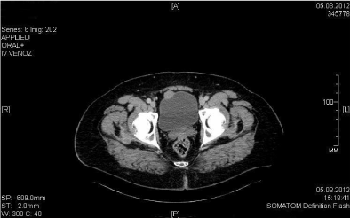
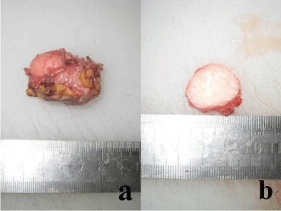
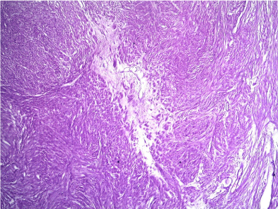
A 55-year old male patient was admitted to Urology department for control ultrasonography (US) due to the chronic prostatitis in history. The US examination revealed a mass at the right anterolateral aspect of the bladder wall. The patient didn’t have any complaints. Subsequent computed tomography (CT) demonstrated well circumscribed soft tissue mass with same localization (Figure 1). On cystoscopy, the existence of a mass with smooth urothelial covering observed at the bladder wall. The mass was resected completely by an open partial cystectomy, under spinal anesthesia. Macroscopically, the specimen was 20×25×30 mm in size with a small amount of surrounding soft tissue, unilaterally mucosa covered, white-gray colored on sectioning and nodular in appearance (Figures 2a, 2b). In histopathological examination of material, tumor was composed mature smooth muscle tissue consisting of spindle cells uniform and elongated (cigar-shaped) nucleuses. There were no atypical features, mitotic figures or coagulative T-cell necrosis (Figure 3). The immunohistochemical study revealed the diffuse positivity with smooth muscle acting in the spindle cells formed the tumor. A case was reported as leimyoma with no special features. The patient was discharged after 3 days with no immediate complications and he was asymptomatic during 25 months follow-up, without any features in control US in the 6th month.
Figure 1 : CT scan shows solid mass on the bladder wall.
Figure 2 : Macroscopic view (a) and cut surface (b) of the resected specimen.
Figure 3 : Microscopic view of tumor tissue contained spindle cells (HE, ×100).
Discussion
Leiomyoma is a rare benign tumor of the urinary bladder, accounting for 0.43% of bladder tumors [4]. Intramural localization of leiomyoma at the bladder wall is rarely seen and constitutes 7% of all cases [5]. The definite etiology of bladder leiomyomas is still uncertain. Chromosomal abnormalities, hormonal influences, bladder musculature infection, perivascular inflammation and dysontogenesis are proposed etiologic factors for bladder leiomyomas [6]. The female predominance at a reproductive age suggests hormonal influence more than the other possibilities.
Bladder leiomyoma usually presents with chronic obstructive or irritative lower urinary tract symptoms. Acute presentation with retention of urine is rare and only few cases have been reported in the literature [2]. However some of the cases did not display any urinary symptoms [7]. Symptoms of bladder leiomyoma are closely related with localization and dimension of the mass. Namely, the endovesical form usually causes irritative or obstructive symptoms or gross hematuria. Intramural and extravesical forms, especially small tumors, may not produce symptoms as in present case. Coexisting bladder and uterine leiomyomas or multiple leiomyomas of the urinary system have been reported [8,9].
Cystoscopy, US, tomography or magnetic resonance imaging (MRI) can be used in the diagnosis, but the definitive diagnosis is made by histopathology.
Usually ultrasonography is the initial imaging modality particularly for incidentally detected leiomyomas, as in our case. Sonographic features of leiomyoma are usually homogeneous hypoechoic, with circumscribed margin and few blood vessels on color Doppler ultrasound. However, the bladder tumors may have variant aspects on imaging evaluation, which may cause misinterpretation and be confused with bladder cancer [10]. On CT, bladder leiomyoma usually presents as a round like hypo dense mass with circumscribed margin; on contrast enhanced CT, it shows centripetal homogeneous enhancement [4]. On MRI, leiomyoma is visualized as low-intensity mass with a smooth surface, while large-degenerative leiomyomas have heterogeneous signal intensity; on contrast enhanced MRI, leiomyomas may show homogeneous enhancement [11]. Cystoscopy is mainly valuable in the diagnostic process of intravesical and symptomatic leiomyomas. Additionally, it is a tool for diagnostic biopsy and transurethral resection.
In the diagnostic process histopathology is of great importance, especially in the differentiation of radiological and cystoscopically suspicious lesions. Small-superficial biopsies taken from surface mucosa for diagnostic purposes can lead to wrong results, especially when present reactive or neoplastic urothelial lesions on the surface [3]. Deep-incisional biopsies can solve this challenge. Should be noted that a rare histological types of leiomyomas as an epithelioid variant also can be seen in the bladder [12].
Treatment of leiomyoma of the urinary bladder is mainly surgical. Surgical options depend on size and location of the tumor, and include transurethral resection of the tumor and open surgical excision [13]. Transurethral resection is performed usually for smaller and endovesical tumors, and it is carried both for therapeutic and diagnostic purposes [3]. However, intramural and extravesical and/ or large leiomyomas are treated by partial cystectomy or enucleation. For this purpose, laparoscopic, transvesical and robotic surgery techniques may be used successfully [8,13,14].
Conclusion
The complete excision of the mass is an important stage in the management of asymptomatic bladder leiomyomas, in order to rule out malignancies. Definitive diagnosis is possible only by histopathological examination. Leiomyoma should be main diagnosis in the differentiation of solid masses of bladder wall.
Acknowledgement
We would like to thank Jeyhun Jalilov and Bahadur Ibrahimov for aid.
References
- Fasih N, Prasad Shanbhogue AK, Macdonald DB, Fraser-Hill MA, Papadatos D, Kielar AZ, et al. Leiomyomas beyond the uterus: unusual locations, rare manifestations. Radiographics. 2008; 28: 1931-1948.
- Agrawal SK, Agrawal P, Paliwal S, Yadav C. Bladder neck leiomyoma presenting with acute retention of urine in an elderly female. J Midlife Health. 2014; 5: 45-48.
- Mammadov R, Musayev J, Hasanov A. Endovesical leiomyoma of bladder: a case report. Georgian Med News. 2013; 7-10.
- Blasco Casares FJ, Sacristán Sanfelipe J, Ibarz Servio L, Batalla Cadira JL, Ruiz Marcellán FJ. [Characteristics of bladder leiomyoma in our setting]. Arch Esp Urol. 1995; 48: 987-990.
- Park JW, Jeong BC, Seo SI, Jeon SS, Kwon GY, Lee HM. Leiomyoma of the urinary bladder: a series of nine cases and review of the literature. Urology. 2010; 76: 1425-1429.
- Cornella JL, Larson TR, Lee RA, Magrina JF, Kammerer-Doak D. Leiomyoma of the female urethra and bladder: report of twenty-three patients and review of the literature. Am J Obstet Gynecol. 1997; 176: 1278-1285.
- Erdem H, Yildirim U, Tekin A, Kayikci A, Uzunlar AK, Sahiner C. Leiomyoma of the urinary bladder in asymptomatic women. Urol Ann. 2012; 4: 172-174.
- Ghadian A, Hoseini SY. Transvesical enucleation of multiple leiomyoma of bladder and urethra. Nephrourol Mon. 2013; 5: 709-711.
- Jain SK, Tanwar R, Mitra A. Bladder Leiomyoma Presenting With LUTS and Coexisting Bladder and Uterine Leiomyomata: A Review of Two Cases. Rev Urol. 2014; 16: 50-54.
- Wu S. Imaging findings of atypical leiomyoma of the urinary bladder simulating bladder cancer: a case report and literature review. Med Ultrason. 2013; 15: 161-163.
- Chen M, Lipson SA, Hricak H. MR imaging evaluation of benign mesenchymal tumors of the urinary bladder. AJR Am J Roentgenol. 1997; 168: 399-403.
- Kaviani A, Razi A, Mokhtarpour H, Mazloomfard MM, Moeini A, Bahrami-Motlagh H. Epitheliod leiomyoma of the bladder: an unusual case of irritative and obstructive voiding symptoms. Case Rep Urol. 2012; 2012: 759150.
- Singh O, Gupta SS, Hastir A. Laparoscopic enucleation of leiomyoma of the urinary bladder: a case report and review of the literature. Urol J. 2011; 8: 155-158.
- Al-Othman KE, Rajih ES, Al-Otaibi MF. Robotic extramucosal excision of bladder wall leiomyoma. Int Braz J Urol. 2014; 40: 127-128.


