Case Report
Austin J Urol. 2014;1(3): 2.
Secondary Vesical Calculus in Female
Chawla A, Mishra DK* and Kumar SP
Department of Urology, Kasturba Medical College, Manipal, Karnataka, INDIA
*Corresponding author: Dr. Mishra DK, Assistant Professor, Department of Urology, KMC, Manipal, Karnataka, INDIA – 576104
Received: August 14, 2014; Accepted: September 22, 2014; Published: September 25, 2014
Abstract
Bladder stones are rarely found in females (2%) and their occurrence should be evaluated in detail. We report an uncommon case of a 64 year old female presenting with calculus over suture in bladder following prior abdominal hysterectomy. The stone was fragmented and removed. An innovative method utilizing a 5 mm laparoscopic port inserted intravesically from suprapubic region and laparoscopic cold scissors was used to pull and cut the suture at its base under cystoscopic guidance.
Keywords: Secondary; Vesical; Calculus: Female
Introduction
Bladder stones are rarely found in females (2%) and their occurrence should be evaluated in detail [1]. The common causes include foreign body or material in bladder resulting from iatrogenic interventions including sling procedures and migrated intrauterine devices [2]. We hereby report an uncommon case of a 64 year old female presenting with calculus over suture in bladder following prior abdominal hysterectomy. An innovative approach of its removal is highlighted.
Case Report
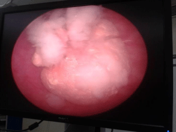
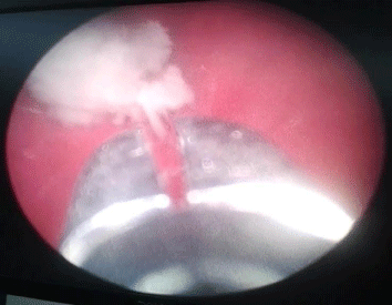
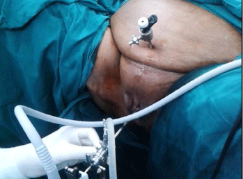
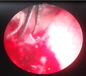
A 64 year old female presented with severe lower urinary tract symptoms (LUTS) since 1 year comprising of frequency and dysuria with occasional debris in urine. She had undergone abdominal hysterectomy for fibroid uterus and menorrhagia 4 years back. Urine examination showed pyuria and microscopic hematuria and urine culture showed E.coli. Ultrasonography revealed a vesical calculus of 2 cm size with some hyperechoic intraluminal lesion in base of bladder. Abdominal roentgenogram was inconclusive. Cystoscopy revealed a 2.5 cm vesical calculus (Figure 1) freely moving and some whitish fluffy material (Figure 2) arising from the base of bladder. The stone was fragmented and removed and revealed to have formed over a suture material (Figure 2). The fluffy material was manipulated and removed and sent for fungal culture which came positive for asperillus flavus. After removal of fluffy material, a white colored suture material was revealed arising from bladder base which on manipulation showed significant indenting of vaginal vault as felt during per vaginal examination. A gentle attempt to remove the suture material by holding with forceps failed. A 5 mm laparoscopic port was inserted intravesically with ultrasonography guidance, from suprapubic region (Figure 3) and laparoscopic cold scissors was used to pull and cut the suture at its base under cystoscopic guidance (Figure 4). Cystogram with instillation of saline mixed with diluted methylene blue dye in bladder did not reveal any leakage per vaginum. Vaginoscopy did not reveal any fistula formation or suture material in vault. On follow up at 1 month, the patient was asymptomatic and urine culture was sterile. Cystoscopy revealed that the site of suture had completely healed.
Figure 1 : Cystoscopy picture of vesical calculus.
Figure 2 : Whitish fluffy material arising from the bladder base over the suture material.
Figure 3 : Suprapubic 5 mm port inserted in bladder with a cystoscope for guidance.
Figure 4 : Suture being divided with cold scissors through the laparoscopic port intravesically.
Discussion
Females account for only 2% of all patients with bladder stones caused by bladder outlet obstruction [1]. The overall incidence of bladder calculi has increased slightly for females over the last 4 decades [3]. This might be due to an increase in the elderly population as well as an overall increase in the number of female genitourinary procedures performed annually [3]. The presentation can comprise of severe LUTS, recurrent UTI (Urinary Tract Infections), suprapubic pain and hematuria.
Animal experiments have revealed that non-absorbable sutures, such as silk and Mersilene, cause substantial tissue reactions. Stones are formed when these sutures are exposed in the bladder cavity [3-4]. Evidence in literature classifies bladder stone formation among females into extra cystic objects such as intrauterine devices which moved ectopically into the bladder [5], intracystic equipment like catheters or sutures left in the bladder [6,7] and bladder damage during surgery with objects such as sutures left embedded in the bladder wall [8,9]. The majority of bladder calculi secondary to female pelvic surgery or after anti-incontinence procedures result from either obstruction or foreign bodies [3]. In our patient, the stone was a result of abdominal hysterectomy done previously which has never been reported. This probably may be due to involvement of bladder wall during vaginal vault closure with non-absorbable suture material. These types of bladder stones are fixed on the bladder wall by the suture and are difficult to remove conventionally. Mechanical crushing and laser fulguration does not remove the suture effectively. Clean division of the suture at the base of the bladder wall is important. This can be achieved effectively by excision of suture via a small 5 mm laparoscopic port inserted suprapubically.
The main principle for managing a bladder stone is to remove the underlying cause of stone formation, such as bladder outlet obstruction or bladder infection [10]. A bladder stone resulting from a foreign body that has become fixed on the bladder wall may occasionally require a laparotomy for its removal [4]. We describe an innovative way to remove the suture fixed to the bladder wall after endoscopic debulking of the stone. A single 5 mm intravesical port placed suprapubically gives advantage in manipulating the suture for its complete removal and can be removed at the end of the procedure with no secondary drainage in form of supra-pubic catheter. However one should be careful in using energy based cutting of the suture as it may lead to an iatrogenic vesico-vaginal fistula (VVF) afterwards.
It is imperative that women who present with significant lower urinary-tract symptoms for longer duration with prior history of urogynecologic procedures, undergo a thorough evaluation to rule out inadvertent intravesical suture placement [3].
Conclusion
The incidence of vesical calculi is on a rise in women due to an increase in pelvic procedures and anti-incontinence surgery. Any female presenting with LUTS and recurrent UTI and vesical calculi should be thoroughly evaluated. Vesical calculi can rarely be caused after abdominal hysterectomy. Suprapubic intravesical insertion of laparoscopic port under cystoscopic guidance offers a new innovation in clean and complete removal of suture material from bladder wall; however one should be careful not to create an iatrogenic VVF in such cases.
Acknowledgements
The authors report no conflicts of interest and no funding was received for this case report.
Authors’ Contributions
DKM, PKS and AC were involved in patient management; DKM and PKS were involved in manuscript preparation; AC reviewed the manuscript and did final approval.
References
- Drach GW. Urinary lithiasis: etiology, diagnosis, and medical treatment. In: Walsh RF, Retik AB, Stamey TA, Vaughan ED (eds). Campbel’’s Urology. 6th (Edn). Philadelphia, PA: WB Saunders Co; 1992: 2085– 2156.
- Benway B M,Bhayani S B. Chapter 89; lower urinary tract calculi. In: Wein A J. Kavoussi L R,Partin A W, Novick A C, Peters C A. (eds) Campbell-Walsh Urology.10th (Edn). Philadelphia, Saunders-Elsevier Inc. 2012: 2523.
- Schwartz BF1, Stoller ML. The vesical calculus. Urol Clin North Am. 2000; 27: 333-346.
- Sheng-Tsun Su, He-Fu Huang, Shu-Fen Chang. Encrusted Bladder Stone on Non-absorbable Sutures after a Cesarean Section: A Case Report. JTUA 2009; 20: 143-145.
- Chamary VL. An unusual cause of iatrogenic bladder stone. Br J Urol. 1995; 76: 138.
- Williams DJ. Uric acid bladder stone associated with a foreign body. Postgrad Med J. 1986; 62: 495.
- Lipke M1, Schulsinger D, Sheynkin Y, Frischer Z, Waltzer W. Endoscopic treatment of bladder calculi in post-renal transplant patients: a 10-year experience. J Endourol. 2004; 18: 787-790.
- Kouriefs C1, Jones CR. Bladder stone following Stamey's colposuspension. Aust N Z J Obstet Gynaecol. 2002; 42: 305-306.
- Zderic SA1, Burros HM, Hanno PM, Dudas N, Whitmore KE. Bladder calculi in women after urethrovesical suspension. J Urol. 1988; 139: 1047-1048.
- Huang WC1, Yang JM. Sonographic appearance of a bladder calculus secondary to a suture from a bladder neck suspension. J Ultrasound Med. 2002; 21: 1303-1305.


